 Movie
Movie Controller

[English] 日本語
 Yorodumi
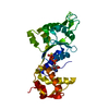
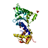
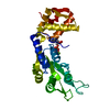
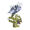
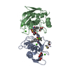
- PDB-6j7e: Crystal Structure of Central domain of FleQ in complex with ATPgS... -
+ Open data

- Basic information
| Entry | Database: PDB / ID: 6j7e | ||||||
|---|---|---|---|---|---|---|---|
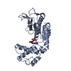
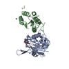
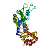
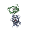
| Title | Crystal Structure of Central domain of FleQ in complex with ATPgS and Mg | ||||||
 Components Components | Nitrogen assimilation regulatory protein | ||||||
 Keywords Keywords | TRANSCRIPTION / FleQ / Pseudomonas / AAA+ / NtrC | ||||||
| Function / homology |  Function and homology information Function and homology informationpositive regulation of cilium-dependent cell motility / regulation of bacterial-type flagellum-dependent cell motility / cyclic-di-GMP binding / positive regulation of cell-substrate adhesion / negative regulation of extracellular matrix assembly / DNA-binding transcription repressor activity / DNA-binding transcription activator activity / cis-regulatory region sequence-specific DNA binding / protein-DNA complex / sequence-specific DNA binding ...positive regulation of cilium-dependent cell motility / regulation of bacterial-type flagellum-dependent cell motility / cyclic-di-GMP binding / positive regulation of cell-substrate adhesion / negative regulation of extracellular matrix assembly / DNA-binding transcription repressor activity / DNA-binding transcription activator activity / cis-regulatory region sequence-specific DNA binding / protein-DNA complex / sequence-specific DNA binding / transcription cis-regulatory region binding / regulation of DNA-templated transcription / positive regulation of DNA-templated transcription / DNA-templated transcription / ATP binding Similarity search - Function | ||||||
| Biological species |   Pseudomonas aeruginosa (bacteria) Pseudomonas aeruginosa (bacteria) | ||||||
| Method |  X-RAY DIFFRACTION / SYNCHROTRON / MOLECULAR REPLACEMENT / Resolution: 2.4 Å X-RAY DIFFRACTION / SYNCHROTRON / MOLECULAR REPLACEMENT / Resolution: 2.4 Å | ||||||
 Authors Authors | Banerjee, P. / Chanchal / Jain, D. | ||||||
| Funding support |  India, 1items India, 1items
| ||||||
 Citation Citation | Journal: Acs Chem.Biol. / Year: 2019 Title: Sensor I Regulated ATPase Activity of FleQ Is Essential for Motility to Biofilm Transition inPseudomonas aeruginosa. Authors: Banerjee, P. / Chanchal / Jain, D. | ||||||
| History |
|
- Structure visualization
| Structure viewer | Molecule: MolmilJmol/JSmol |
|---|
- Downloads & links
-Download
| PDBx/mmCIF format | 6j7e.cif.gz | 69.3 KB | Display | PDBx/mmCIF format |
|---|---|---|---|---|
| PDB format | pdb6j7e.ent.gz | 48.3 KB | Display | PDB format |
| PDBx/mmJSON format | 6j7e.json.gz | Tree view | PDBx/mmJSON format | |
| Others |  Other downloads Other downloads |
-Validation report
| Arichive directory | https://data.pdbj.org/pub/pdb/validation_reports/j7/6j7eftp://data.pdbj.org/pub/pdb/validation_reports/j7/6j7e | HTTPS FTP |
|---|
-Related structure data
| Related structure data |  6jdiC  6jdlC  5expS S: Starting model for refinement C: citing same article ( |
|---|---|
| Similar structure data |

-Links
 PDBj
PDBj


- Assembly
| Deposited unit | 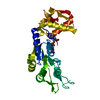
| ||||||||
|---|---|---|---|---|---|---|---|---|---|
| 1 |
| ||||||||
| Unit cell |
|
-Components
| #1: Protein | Mass: 29599.936 Da / Num. of mol.: 1 / Fragment: Central Domain Source method: isolated from a genetically manipulated source Source: (gene. exp.) Pseudomonas aeruginosa (bacteria)Gene: ntrC_2, fleQ, ntrC_1, C8257_22345, CAZ10_06755, CGU42_27830, DZ940_07790, DZ962_01740, NCTC13719_04150, PAERUG_E15_London_28_01_14_04909, PAMH19_4438, RW109_RW109_05080 Production host: |
|---|---|
| #2: Chemical | ChemComp-AGS /   Mass: 523.247 Da / Num. of mol.: 1 / Source method: obtained synthetically / Formula: C10H16N5O12P3S / Comment: ATP-gamma-S, energy-carrying molecule analogue*YM Mass: 523.247 Da / Num. of mol.: 1 / Source method: obtained synthetically / Formula: C10H16N5O12P3S / Comment: ATP-gamma-S, energy-carrying molecule analogue*YM |
| #3: Chemical | ChemComp-MG /   Mass: 24.305 Da / Num. of mol.: 1 / Source method: obtained synthetically / Formula: Mg Mass: 24.305 Da / Num. of mol.: 1 / Source method: obtained synthetically / Formula: Mg |
| #4: Chemical | ChemComp-GOL /   Mass: 92.094 Da / Num. of mol.: 1 / Source method: obtained synthetically / Formula: C3H8O3 Mass: 92.094 Da / Num. of mol.: 1 / Source method: obtained synthetically / Formula: C3H8O3 |
| #5: Water | ChemComp-HOH /  Mass: 18.015 Da / Num. of mol.: 70 / Source method: isolated from a natural source / Formula: H2O Mass: 18.015 Da / Num. of mol.: 70 / Source method: isolated from a natural source / Formula: H2O |
-Experimental details
-Experiment
| Experiment | Method: X-RAY DIFFRACTION / Number of used crystals: 1 |
|---|
- Sample preparation
| Crystal | Density Matthews: 2.28 Å3/Da / Density % sol: 46.03 % |
|---|---|
| Crystal grow | Temperature: 281 K / Method: vapor diffusion, hanging drop / Details: 0.2M of sodium thiocyanate, 22% of PEG 3350 |
-Data collection
| Diffraction | Mean temperature: 100 K / Serial crystal experiment: N |
|---|---|
| Diffraction source | Source: SYNCHROTRON / Site: ESRF  / Beamline: ID29 / Wavelength: 0.97319 Å / Beamline: ID29 / Wavelength: 0.97319 Å |
| Detector | Type: DECTRIS PILATUS 6M / Detector: PIXEL / Date: Mar 17, 2017 |
| Radiation | Protocol: SINGLE WAVELENGTH / Monochromatic (M) / Laue (L): M / Scattering type: x-ray |
| Radiation wavelength | Wavelength: 0.97319 Å / Relative weight: 1 |
| Reflection | Resolution: 2.4→90.6 Å / Num. obs: 10591 / % possible obs: 99.4 % / Redundancy: 3.6 % / Rmerge(I) obs: 0.096 / Net I/σ(I): 9.7 |
| Reflection shell | Resolution: 2.4→2.49 Å / Rmerge(I) obs: 0.663 |
- Processing
| Software |
| |||||||||||||||||||||||||||||||||||
|---|---|---|---|---|---|---|---|---|---|---|---|---|---|---|---|---|---|---|---|---|---|---|---|---|---|---|---|---|---|---|---|---|---|---|---|---|
| Refinement | Method to determine structure: MOLECULAR REPLACEMENT Starting model: 5EXP Resolution: 2.4→52.274 Å / SU ML: 0.36 / Cross valid method: FREE R-VALUE / σ(F): 1.34 / Phase error: 27.82 / Stereochemistry target values: ML
| |||||||||||||||||||||||||||||||||||
| Solvent computation | Shrinkage radii: 0.9 Å / VDW probe radii: 1.11 Å / Solvent model: FLAT BULK SOLVENT MODEL | |||||||||||||||||||||||||||||||||||
| Refinement step | Cycle: LAST / Resolution: 2.4→52.274 Å
| |||||||||||||||||||||||||||||||||||
| Refine LS restraints |
| |||||||||||||||||||||||||||||||||||
| LS refinement shell |
|
