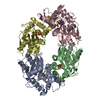
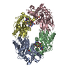
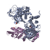
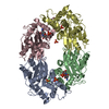
登録情報 データベース : PDB / ID : 6j6uタイトル Rat PTPRZ D1-D2 domain Receptor-type tyrosine-protein phosphatase zeta キーワード / 機能・相同性 分子機能 ドメイン・相同性 構成要素
/ / / / / / / / / / / / / / / / / / / / / / / / / / / / / / / / / / / / / / / / / / / / / / / / / / / / / / / / / / / / / / / / / / / / / / / / / / / / / / / / / / / / / / / / / / / 生物種 Rattus norvegicus (ドブネズミ)手法 / / 解像度 : 3.32 Å データ登録者 Sugawara, H. #1: ジャーナル : Scientific Reports / 年 : 2016タイトル : Small-molecule inhibition of PTPRZ suppresses tumor growth in a rat model of glioblastoma.
著者 :
Fujikawa, A. / Nagahira, A. / Sugawara, H. / Ishii, K. / Imajo, S. / Matsumoto, M. / Kuboyama, K. / Suzuki, R. / Tanga, N. / Noda, M. / Uchiyama, S. / Tomoo, T. / Ogata, A. / Masumura, M. / Noda, M. #2: ジャーナル : Sci Rep / 年 : 2017タイトル : Targeting PTPRZ inhibits stem cell-like properties and tumorigenicity in glioblastoma cells.
著者 :
Fujikawa, A. / Sugawara, H. / Tanaka, T. / Matsumoto, M. / Kuboyama, K. / Suzuki, R. / Tanga, N. / Ogata, A. / Masumura, M. / Noda, M. 履歴 登録 2019年1月15日 登録サイト / 処理サイト 改定 1.0 2019年8月28日 Provider / タイプ 改定 1.1 2019年10月30日 Group / Database references / カテゴリ Item / _citation.page_first / _citation.page_last改定 1.2 2023年11月22日 Group / Database references / Refinement descriptionカテゴリ chem_comp_atom / chem_comp_bond ... chem_comp_atom / chem_comp_bond / database_2 / pdbx_initial_refinement_model / struct_ncs_dom_lim Item _database_2.pdbx_DOI / _database_2.pdbx_database_accession ... _database_2.pdbx_DOI / _database_2.pdbx_database_accession / _struct_ncs_dom_lim.beg_auth_comp_id / _struct_ncs_dom_lim.beg_label_asym_id / _struct_ncs_dom_lim.beg_label_comp_id / _struct_ncs_dom_lim.beg_label_seq_id / _struct_ncs_dom_lim.end_auth_comp_id / _struct_ncs_dom_lim.end_label_asym_id / _struct_ncs_dom_lim.end_label_comp_id / _struct_ncs_dom_lim.end_label_seq_id
すべて表示 表示を減らす
 ムービー
ムービー コントローラー


 データを開く
 基本情報
基本情報 要素
要素 キーワード
キーワード 機能・相同性情報
 X線回折 /
  データ登録者
 引用
引用 構造の表示
構造の表示 ダウンロードとリンク
 その他のダウンロード


 PDBj
PDBj


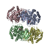
 集合体
集合体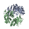

 試料調製
試料調製 解析
解析