 Movie
Movie Controller

[English] 日本語
 Yorodumi
- PDB-6j5u: Ligand-triggered allosteric ADP release primes a plant NLR complex -
+ Open data

- Basic information
| Entry | Database: PDB / ID: 6j5u | |||||||||
|---|---|---|---|---|---|---|---|---|---|---|
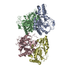
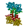
| Title | Ligand-triggered allosteric ADP release primes a plant NLR complex | |||||||||
 Components Components |
| |||||||||
 Keywords Keywords | PLANT PROTEIN / ZAR1 / RKS1 / PBL2-UMP | |||||||||
| Function / homology |  Function and homology information Function and homology informationpositive regulation of defense response to bacterium / Tat protein binding / response to temperature stimulus / regulation of immune response / defense response / ADP binding / kinase activity / defense response to Gram-negative bacterium / eukaryotic translation initiation factor 2alpha kinase activity / 3-phosphoinositide-dependent protein kinase activity ...positive regulation of defense response to bacterium / Tat protein binding / response to temperature stimulus / regulation of immune response / defense response / ADP binding / kinase activity / defense response to Gram-negative bacterium / eukaryotic translation initiation factor 2alpha kinase activity / 3-phosphoinositide-dependent protein kinase activity / DNA-dependent protein kinase activity / ribosomal protein S6 kinase activity / histone H3S10 kinase activity / histone H2AXS139 kinase activity / histone H3S28 kinase activity / histone H4S1 kinase activity / histone H2BS14 kinase activity / histone H3T3 kinase activity / histone H2AS121 kinase activity / Rho-dependent protein serine/threonine kinase activity / histone H2BS36 kinase activity / histone H3S57 kinase activity / histone H2AT120 kinase activity / AMP-activated protein kinase activity / histone H2AS1 kinase activity / histone H3T6 kinase activity / histone H3T11 kinase activity / histone H3T45 kinase activity / cell surface receptor signaling pathway / non-specific serine/threonine protein kinase / defense response to bacterium / protein serine kinase activity / ATP binding / nucleus / plasma membrane Similarity search - Function | |||||||||
| Biological species |  | |||||||||
| Method | ELECTRON MICROSCOPY / single particle reconstruction / cryo EM / Resolution: 3.9 Å | |||||||||
 Authors Authors | Wang, J.Z. / Wang, J. / Meijuan, H. / Wang, H.W. / Zhou, J.M. / Chai, J.J. | |||||||||
| Funding support |  China, 2items China, 2items
| |||||||||
 Citation Citation | Journal: Science / Year: 2019 Title: Ligand-triggered allosteric ADP release primes a plant NLR complex. Authors: Jizong Wang / Jia Wang / Meijuan Hu / Shan Wu / Jinfeng Qi / Guoxun Wang / Zhifu Han / Yijun Qi / Ning Gao / Hong-Wei Wang / Jian-Min Zhou / Jijie Chai /  Abstract: Pathogen recognition by nucleotide-binding (NB), leucine-rich repeat (LRR) receptors (NLRs) plays roles in plant immunity. The pv. effector AvrAC uridylylates the PBL2 kinase, and the latter (PBL2) ...Pathogen recognition by nucleotide-binding (NB), leucine-rich repeat (LRR) receptors (NLRs) plays roles in plant immunity. The pv. effector AvrAC uridylylates the PBL2 kinase, and the latter (PBL2) acts as a ligand to activate the NLR ZAR1 precomplexed with the RKS1 pseudokinase. Here we report the cryo-electron microscopy structures of ZAR1-RKS1 and ZAR1-RKS1-PBL2 in an inactive and intermediate state, respectively. The ZAR1 domain, compared with animal NLR domains, is differently positioned to sequester ZAR1 in an inactive state. Recognition of PBL2 is exclusively through RKS1, which interacts with ZAR1 PBL2 binding stabilizes the RKS1 activation segment, which sterically blocks ZAR1 adenosine diphosphate (ADP) binding. This engenders a more flexible NB domain without conformational changes in the other ZAR1 domains. Our study provides a structural template for understanding plant NLRs. | |||||||||
| History |
|
- Structure visualization
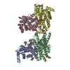
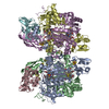
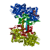
| Movie |
Movie viewer |
|---|---|
| Structure viewer | Molecule: MolmilJmol/JSmol |
- Downloads & links
-Download
| PDBx/mmCIF format | 6j5u.cif.gz | 228 KB | Display | PDBx/mmCIF format |
|---|---|---|---|---|
| PDB format | pdb6j5u.ent.gz | 173.5 KB | Display | PDB format |
| PDBx/mmJSON format | 6j5u.json.gz | Tree view | PDBx/mmJSON format | |
| Others |  Other downloads Other downloads |
-Validation report
| Arichive directory | https://data.pdbj.org/pub/pdb/validation_reports/j5/6j5uftp://data.pdbj.org/pub/pdb/validation_reports/j5/6j5u | HTTPS FTP |
|---|
-Related structure data
| Related structure data |  0681MC 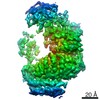 0682C  0683C  6j5vC  6j5wC M: map data used to model this data C: citing same article ( |
|---|---|
| Similar structure data |


-Links
 PDBj
PDBj


- Assembly
| Deposited unit | 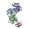
|
|---|---|
| 1 |
|
-Components
| #1: Protein | Mass: 97163.977 Da / Num. of mol.: 1 Source method: isolated from a genetically manipulated source Source: (gene. exp.) Production host: Insect cell expression vector pTIE1 (others) References: UniProt: Q38834 | ||
|---|---|---|---|
| #2: Protein | Mass: 46356.430 Da / Num. of mol.: 1 Source method: isolated from a genetically manipulated source Source: (gene. exp.) Production host: Insect cell expression vector pTIE1 (others) References: UniProt: O49839, non-specific serine/threonine protein kinase | ||
| #3: Protein | Mass: 40142.375 Da / Num. of mol.: 1 Source method: isolated from a genetically manipulated source Source: (gene. exp.) Gene: F15B8.100, Resistance related KinaSe 1, RKS1, At3g57710 Production host: Insect cell expression vector pTIE1 (others) References: UniProt: Q9SVY5 | ||
| #4: Chemical |   Mass: 324.181 Da / Num. of mol.: 2 / Source method: obtained synthetically / Formula: C9H13N2O9P Mass: 324.181 Da / Num. of mol.: 2 / Source method: obtained synthetically / Formula: C9H13N2O9PHas protein modification | Y | |
-Experimental details
-Experiment
| Experiment | Method: ELECTRON MICROSCOPY |
|---|---|
| EM experiment | Aggregation state: PARTICLE / 3D reconstruction method: single particle reconstruction |
- Sample preparation
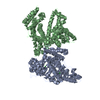
| Component | Name: ZAR1-RKS1 with ADP complex / Type: COMPLEX / Entity ID: #1 / Source: RECOMBINANT |
|---|---|
| Molecular weight | Value: 0.13 MDa / Experimental value: YES |
| Source (natural) | Organism: |
| Source (recombinant) | Organism: Insect cell expression vector pTIE1 (others) |
| Buffer solution | pH: 8 |
| Specimen | Embedding applied: NO / Shadowing applied: NO / Staining applied: NO / Vitrification applied: YES |
| Vitrification | Instrument: FEI VITROBOT MARK IV / Cryogen name: ETHANE / Humidity: 100 % / Chamber temperature: 295 K |
- Electron microscopy imaging
| Experimental equipment |  Model: Titan Krios / Image courtesy: FEI Company |
|---|---|
| Microscopy | Model: FEI TITAN KRIOS |
| Electron gun | Electron source:  FIELD EMISSION GUN / Accelerating voltage: 300 kV / Illumination mode: FLOOD BEAM FIELD EMISSION GUN / Accelerating voltage: 300 kV / Illumination mode: FLOOD BEAM |
| Electron lens | Mode: BRIGHT FIELD / Nominal magnification: 105000 X / Nominal defocus max: 900 nm / Nominal defocus min: 700 nm / Cs: 0.01 mm / C2 aperture diameter: 50 µm |
| Specimen holder | Cryogen: NITROGEN / Specimen holder model: FEI TITAN KRIOS AUTOGRID HOLDER |
| Image recording | Electron dose: 50 e/Å2 / Film or detector model: GATAN K2 SUMMIT (4k x 4k) |
- Processing
| Software | Name: PHENIX / Version: 1.13_2998: / Classification: refinement | ||||||||||||||||||
|---|---|---|---|---|---|---|---|---|---|---|---|---|---|---|---|---|---|---|---|
| EM software |
| ||||||||||||||||||
| CTF correction | Type: PHASE FLIPPING AND AMPLITUDE CORRECTION | ||||||||||||||||||
| Particle selection | Num. of particles selected: 3342829 | ||||||||||||||||||
| Symmetry | Point symmetry: C1 (asymmetric) | ||||||||||||||||||
| 3D reconstruction | Resolution: 3.9 Å / Num. of particles: 404311 / Symmetry type: POINT | ||||||||||||||||||
| Atomic model building | Space: REAL | ||||||||||||||||||
| Atomic model building | PDB-ID: 3TL8 Accession code: 3TL8 / Source name: PDB / Type: experimental model | ||||||||||||||||||
| Refinement | Highest resolution: 3.9 Å |

