 Movie
Movie Controller


+ Open data

- Basic information
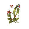
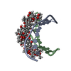
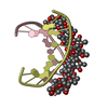
| Entry | Database: PDB / ID: 6j0i | |||||||||||||||||||||||
|---|---|---|---|---|---|---|---|---|---|---|---|---|---|---|---|---|---|---|---|---|---|---|---|---|
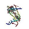
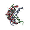
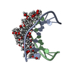
| Title | Structure of [Co2+-(Chromomycin A3)2]-d(TTGGCGAA)2 complex | |||||||||||||||||||||||
 Components Components | DNA (5'-D(* Keywords KeywordsDNA/ANTIBIOTIC / Mismatch DNA / Chromomycin A3 / Drug-DNA complex / G:G mismatch / DNA-ANTIBIOTIC complex | Function / homology | : / Chem-CRH / DNA |  Function and homology information Function and homology informationBiological species | synthetic construct (others) | Method |  X-RAY DIFFRACTION / SYNCHROTRON / MOLECULAR REPLACEMENT / Resolution: 2.5 Å X-RAY DIFFRACTION / SYNCHROTRON / MOLECULAR REPLACEMENT / Resolution: 2.5 Å  Authors AuthorsSatange, R.B. / Chuang, C.Y. / Hou, M.H. |  CitationJournal: Nucleic Acids Res. / Year: 2019 CitationJournal: Nucleic Acids Res. / Year: 2019Title: Polymorphic G:G mismatches act as hotspots for inducing right-handed Z DNA by DNA intercalation. Authors: Satange, R. / Chuang, C.Y. / Neidle, S. / Hou, M.H. History |
|
- Structure visualization
| Structure viewer | Molecule: MolmilJmol/JSmol |
|---|
- Downloads & links
-Download
| PDBx/mmCIF format | 6j0i.cif.gz | 63.8 KB | Display | PDBx/mmCIF format |
|---|---|---|---|---|
| PDB format | pdb6j0i.ent.gz | 46.7 KB | Display | PDB format |
| PDBx/mmJSON format | 6j0i.json.gz | Tree view | PDBx/mmJSON format | |
| Others |  Other downloads Other downloads |
-Validation report
| Summary document | 6j0i_validation.pdf.gz | 4.6 MB | Display | wwPDB validaton report |
|---|---|---|---|---|
| Full document | 6j0i_full_validation.pdf.gz | 4.6 MB | Display | |
| Data in XML | 6j0i_validation.xml.gz | 17 KB | Display | |
| Data in CIF | 6j0i_validation.cif.gz | 23.5 KB | Display | |
| Arichive directory | https://data.pdbj.org/pub/pdb/validation_reports/j0/6j0iftp://data.pdbj.org/pub/pdb/validation_reports/j0/6j0i | HTTPS FTP |
-Related structure data
| Related structure data |  6j0hC 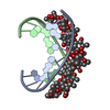 1vaqS S: Starting model for refinement C: citing same article ( |
|---|---|
| Similar structure data |


-Links
 PDBj
PDBj
- Assembly
| Deposited unit | 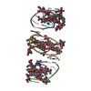
| |||||||||||||||
|---|---|---|---|---|---|---|---|---|---|---|---|---|---|---|---|---|
| 1 | 
| |||||||||||||||
| 2 | 
| |||||||||||||||
| 3 | 
| |||||||||||||||
| Unit cell |
| |||||||||||||||
| Components on special symmetry positions |
|
-Components
-DNA chain , 1 types, 6 molecules ABCDEF
| #1: DNA chain | Mass: 2466.641 Da / Num. of mol.: 6 / Source method: obtained synthetically / Source: (synth.) synthetic construct (others) |
|---|
-Sugars , 2 types, 12 molecules
| #2: Polysaccharide | 2,6-dideoxy-4-O-methyl-alpha-D-galactopyranose-(1-3)-(2R,3R,6R)-6-hydroxy-2-methyltetrahydro-2H- ...2,6-dideoxy-4-O-methyl-alpha-D-galactopyranose-(1-3)-(2R,3R,6R)-6-hydroxy-2-methyltetrahydro-2H-pyran-3-yl acetate Source method: isolated from a genetically manipulated source #3: Polysaccharide | 3-C-methyl-4-O-acetyl-alpha-L-Olivopyranose-(1-3)-(2R,5S,6R)-6-methyltetrahydro-2H-pyran-2,5-diol- ...3-C-methyl-4-O-acetyl-alpha-L-Olivopyranose-(1-3)-(2R,5S,6R)-6-methyltetrahydro-2H-pyran-2,5-diol-(1-3)-(2R,5S,6R)-6-methyltetrahydro-2H-pyran-2,5-diol Source method: isolated from a genetically manipulated source |
|---|
-Non-polymers , 3 types, 394 molecules 


| #4: Chemical | ChemComp-CRH /  Mass: 388.411 Da / Num. of mol.: 6 Mass: 388.411 Da / Num. of mol.: 6Source method: isolated from a genetically manipulated source Formula: C21H24O7 #5: Chemical | ChemComp-CO /  Mass: 58.933 Da / Num. of mol.: 7 Mass: 58.933 Da / Num. of mol.: 7Source method: isolated from a genetically manipulated source Formula: Co #6: Water | ChemComp-HOH / | Mass: 18.015 Da / Num. of mol.: 381 / Source method: isolated from a natural source / Formula: H2O |
|---|
-Experimental details
-Experiment
| Experiment | Method: X-RAY DIFFRACTION / Number of used crystals: 1 |
|---|
- Sample preparation
| Crystal | Density Matthews: 3.76 Å3/Da / Density % sol: 67.27 % |
|---|---|
| Crystal grow | Temperature: 277.15 K / Method: vapor diffusion, sitting drop / pH: 7.3 Details: 5mM Sodium Cacodylate (pH 7.3), 7mM MgCl2, 12mM Spermine, 4% PEG 400, 4% 1-propanol |
-Data collection
| Diffraction | Mean temperature: 100 K / Serial crystal experiment: N |
|---|---|
| Diffraction source | Source: SYNCHROTRON / Site: SPring-8  / Beamline: BL44XU / Wavelength: 0.9 Å / Beamline: BL44XU / Wavelength: 0.9 Å |
| Detector | Type: Bruker DIP-6040 / Detector: CCD / Date: Nov 12, 2014 |
| Radiation | Protocol: SINGLE WAVELENGTH / Monochromatic (M) / Laue (L): M / Scattering type: x-ray |
| Radiation wavelength | Wavelength: 0.9 Å / Relative weight: 1 |
| Reflection | Resolution: 2.5→20 Å / Num. obs: 12624 / % possible obs: 95.5 % / Redundancy: 6.8 % / Biso Wilson estimate: 39.79 Å2 / Rmerge(I) obs: 0.043 / Rpim(I) all: 0.019 / Rrim(I) all: 0.047 / Χ2: 0.914 / Net I/σ(I): 21.7 / Num. measured all: 53369 |
| Reflection shell | Resolution: 2.5→2.59 Å / Redundancy: 7.2 % / Rmerge(I) obs: 0.512 / Num. unique obs: 549 / CC1/2: 0.996 / Rpim(I) all: 0.017 / Rrim(I) all: 0.037 / Χ2: 0.786 / % possible all: 100 |
- Processing
| Software |
| |||||||||||||||||||||||||||||||||||
|---|---|---|---|---|---|---|---|---|---|---|---|---|---|---|---|---|---|---|---|---|---|---|---|---|---|---|---|---|---|---|---|---|---|---|---|---|
| Refinement | Method to determine structure: MOLECULAR REPLACEMENT Starting model: 1VAQ Resolution: 2.5→16.12 Å / SU ML: 0.46 / Cross valid method: THROUGHOUT / σ(F): 1.35 / Phase error: 31.79
| |||||||||||||||||||||||||||||||||||
| Solvent computation | Shrinkage radii: 0.9 Å / VDW probe radii: 1.11 Å | |||||||||||||||||||||||||||||||||||
| Displacement parameters | Biso max: 62 Å2 / Biso mean: 39.42 Å2 / Biso min: 14.33 Å2 | |||||||||||||||||||||||||||||||||||
| Refinement step | Cycle: final / Resolution: 2.5→16.12 Å
| |||||||||||||||||||||||||||||||||||
| LS refinement shell | Refine-ID: X-RAY DIFFRACTION / Rfactor Rfree error: 0 / Total num. of bins used: 4
|
