[English] 日本語
 Yorodumi
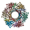
- PDB-6j0b: Cryo-EM Structure of an Extracellular Contractile Injection Syste... -
+ Open data
- Basic information
| Entry | Database: PDB / ID: 6j0b | ||||||
|---|---|---|---|---|---|---|---|
| Title | Cryo-EM Structure of an Extracellular Contractile Injection System, PVC sheath-tube complex in extended state | ||||||
 Components Components |
| ||||||
 Keywords Keywords | PROTEIN TRANSPORT / assembly / Photorhabdus asymbiotica / PVC / contractile injection system / bacteriophage-like | ||||||
| Function / homology |  Function and homology information Function and homology information | ||||||
| Biological species |  Photorhabdus asymbiotica subsp. asymbiotica (bacteria) Photorhabdus asymbiotica subsp. asymbiotica (bacteria) | ||||||
| Method | ELECTRON MICROSCOPY / helical reconstruction / cryo EM / Resolution: 2.9 Å | ||||||
 Authors Authors | Jiang, F. / Li, N. / Wang, X. / Cheng, J. / Huang, Y. / Yang, Y. / Yang, J. / Cai, B. / Wang, Y. / Jin, Q. / Gao, N. | ||||||
 Citation Citation |  Journal: Cell / Year: 2019 Journal: Cell / Year: 2019Title: Cryo-EM Structure and Assembly of an Extracellular Contractile Injection System. Authors: Feng Jiang / Ningning Li / Xia Wang / Jiaxuan Cheng / Yaoguang Huang / Yun Yang / Jianguo Yang / Bin Cai / Yi-Ping Wang / Qi Jin / Ning Gao /  Abstract: Contractile injection systems (CISs) are cell-puncturing nanodevices that share ancestry with contractile tail bacteriophages. Photorhabdus virulence cassette (PVC) represents one group of ...Contractile injection systems (CISs) are cell-puncturing nanodevices that share ancestry with contractile tail bacteriophages. Photorhabdus virulence cassette (PVC) represents one group of extracellular CISs that are present in both bacteria and archaea. Here, we report the cryo-EM structure of an intact PVC from P. asymbiotica. This over 10-MDa device resembles a simplified T4 phage tail, containing a hexagonal baseplate complex with six fibers and a capped 117-nanometer sheath-tube trunk. One distinct feature of the PVC is the presence of three variants for both tube and sheath proteins, indicating a functional specialization of them during evolution. The terminal hexameric cap docks onto the topmost layer of the inner tube and locks the outer sheath in pre-contraction state with six stretching arms. Our results on the PVC provide a framework for understanding the general mechanism of widespread CISs and pave the way for using them as delivery tools in biological or therapeutic applications. | ||||||
| History |
|
- Structure visualization
| Movie |
 Movie viewer Movie viewer |
|---|---|
| Structure viewer | Molecule:  Molmil Molmil Jmol/JSmol Jmol/JSmol |
- Downloads & links
- Download
| PDBx/mmCIF format |  6j0b.cif.gz 6j0b.cif.gz | 970.8 KB | Display |  PDBx/mmCIF format PDBx/mmCIF format |
|---|---|---|---|---|
| PDB format |  pdb6j0b.ent.gz pdb6j0b.ent.gz | 829.9 KB | Display |  PDB format PDB format |
| PDBx/mmJSON format |  6j0b.json.gz 6j0b.json.gz | Tree view |  PDBx/mmJSON format PDBx/mmJSON format | |
| Others |  Other downloads Other downloads |
-Validation report
| Arichive directory |  https://data.pdbj.org/pub/pdb/validation_reports/j0/6j0b https://data.pdbj.org/pub/pdb/validation_reports/j0/6j0b ftp://data.pdbj.org/pub/pdb/validation_reports/j0/6j0b ftp://data.pdbj.org/pub/pdb/validation_reports/j0/6j0b | HTTPS FTP |
|---|
-Related structure data
| Related structure data |  9760MC  9761C  9762C  9763C  9764C  9765C  6j0cC  6j0fC  6j0mC  6j0nC M: map data used to model this data C: citing same article ( |
|---|---|
| Similar structure data |
- Links
Links
- Assembly
| Deposited unit | 
|
|---|---|
| 1 |
|
- Components
| #1: Protein | Mass: 39374.281 Da / Num. of mol.: 12 Source method: isolated from a genetically manipulated source Source: (gene. exp.)  Photorhabdus asymbiotica subsp. asymbiotica (strain ATCC 43949 / 3105-77) (bacteria) Photorhabdus asymbiotica subsp. asymbiotica (strain ATCC 43949 / 3105-77) (bacteria)Strain: ATCC 43949 / 3105-77 / Gene: PAU_03352, PA-RVA20-21-0170 / Production host:  #2: Protein | Mass: 16532.557 Da / Num. of mol.: 12 Source method: isolated from a genetically manipulated source Source: (gene. exp.)  Photorhabdus asymbiotica subsp. asymbiotica (strain ATCC 43949 / 3105-77) (bacteria) Photorhabdus asymbiotica subsp. asymbiotica (strain ATCC 43949 / 3105-77) (bacteria)Strain: ATCC 43949 / 3105-77 / Gene: PAU_03353, PA-RVA20-21-0171 / Production host:  |
|---|
-Experimental details
-Experiment
| Experiment | Method: ELECTRON MICROSCOPY |
|---|---|
| EM experiment | Aggregation state: HELICAL ARRAY / 3D reconstruction method: helical reconstruction |
- Sample preparation
| Component | Name: sheath and tube in extended state / Type: COMPLEX / Entity ID: all / Source: RECOMBINANT |
|---|---|
| Source (natural) | Organism:  Photorhabdus asymbiotica (bacteria) Photorhabdus asymbiotica (bacteria) |
| Source (recombinant) | Organism:  |
| Buffer solution | pH: 7.4 |
| Specimen | Embedding applied: NO / Shadowing applied: NO / Staining applied: NO / Vitrification applied: YES |
| Specimen support | Grid material: COPPER / Grid type: Quantifoil R2/2 |
| Vitrification | Instrument: FEI VITROBOT MARK IV / Cryogen name: ETHANE / Humidity: 100 % |
- Electron microscopy imaging
| Experimental equipment |  Model: Titan Krios / Image courtesy: FEI Company |
|---|---|
| Microscopy | Model: FEI TITAN KRIOS |
| Electron gun | Electron source:  FIELD EMISSION GUN / Accelerating voltage: 300 kV / Illumination mode: FLOOD BEAM FIELD EMISSION GUN / Accelerating voltage: 300 kV / Illumination mode: FLOOD BEAM |
| Electron lens | Mode: BRIGHT FIELD / Alignment procedure: COMA FREE |
| Specimen holder | Cryogen: NITROGEN / Specimen holder model: FEI TITAN KRIOS AUTOGRID HOLDER |
| Image recording | Electron dose: 46.4 e/Å2 / Detector mode: INTEGRATING / Film or detector model: FEI FALCON II (4k x 4k) |
- Processing
| CTF correction | Type: PHASE FLIPPING AND AMPLITUDE CORRECTION |
|---|---|
| Helical symmerty | Angular rotation/subunit: 19.9 ° / Axial rise/subunit: 39.3 Å / Axial symmetry: C6 |
| 3D reconstruction | Resolution: 2.9 Å / Resolution method: FSC 0.143 CUT-OFF / Num. of particles: 551000 / Symmetry type: HELICAL |
 Movie
Movie Controller


 PDBj
PDBj