 Movie
Movie Controller


+ Open data

- Basic information
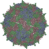
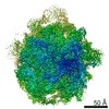
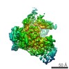
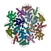
| Entry | Database: PDB / ID: 5cdd | ||||||
|---|---|---|---|---|---|---|---|
| Title | Crystal Structure of Israel acute Paralysis Virus Pentamer | ||||||
 Components Components |
| ||||||
 Keywords Keywords | VIRUS / Pentamer / capsid / honeybee virus / pathogen | ||||||
| Function / homology |  Function and homology information Function and homology information | ||||||
| Biological species |  Israeli acute paralysis virus Israeli acute paralysis virus | ||||||
| Method |  X-RAY DIFFRACTION / SYNCHROTRON / MOLECULAR REPLACEMENT / molecular replacement / Resolution: 2.7 Å X-RAY DIFFRACTION / SYNCHROTRON / MOLECULAR REPLACEMENT / molecular replacement / Resolution: 2.7 Å | ||||||
 Authors Authors | Mullapudi, E. / Plevka, P. | ||||||
 Citation Citation | Journal: J.Virol. / Year: 2016 Title: Virion Structure of Iflavirus Slow Bee Paralysis Virus at 2.6-Angstrom Resolution. Authors: Kalynych, S. / Pridal, A. / Palkova, L. / Levdansky, Y. / de Miranda, J.R. / Plevka, P. | ||||||
| History |
|
- Structure visualization
| Structure viewer | Molecule: MolmilJmol/JSmol |
|---|
- Downloads & links
-Download
| PDBx/mmCIF format | 5cdd.cif.gz | 151.4 KB | Display | PDBx/mmCIF format |
|---|---|---|---|---|
| PDB format | pdb5cdd.ent.gz | 118.4 KB | Display | PDB format |
| PDBx/mmJSON format | 5cdd.json.gz | Tree view | PDBx/mmJSON format | |
| Others |  Other downloads Other downloads |
-Validation report
| Arichive directory | https://data.pdbj.org/pub/pdb/validation_reports/cd/5cddftp://data.pdbj.org/pub/pdb/validation_reports/cd/5cdd | HTTPS FTP |
|---|
-Related structure data


-Links
 PDBj
PDBj
- Assembly
| Deposited unit | 
| ||||||||||||||||||||||||||||||||||||||||||||
|---|---|---|---|---|---|---|---|---|---|---|---|---|---|---|---|---|---|---|---|---|---|---|---|---|---|---|---|---|---|---|---|---|---|---|---|---|---|---|---|---|---|---|---|---|---|
| 1 | x 10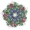
| ||||||||||||||||||||||||||||||||||||||||||||
| 2 |
| ||||||||||||||||||||||||||||||||||||||||||||
| 3 | 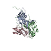
| ||||||||||||||||||||||||||||||||||||||||||||
| Unit cell |
| ||||||||||||||||||||||||||||||||||||||||||||
| Symmetry | Point symmetry: (Schoenflies symbol: D5 (2x5 fold dihedral)) | ||||||||||||||||||||||||||||||||||||||||||||
| Noncrystallographic symmetry (NCS) | NCS oper:
|
-Components
| #1: Protein | Mass: 23881.695 Da / Num. of mol.: 1 / Source method: isolated from a natural source / Source: (natural) Israeli acute paralysis virus / References: UniProt: B3TZF1 |
|---|---|
| #2: Protein | Mass: 33353.836 Da / Num. of mol.: 1 / Source method: isolated from a natural source / Source: (natural) Israeli acute paralysis virus / References: UniProt: D1FK67 |
| #3: Protein | Mass: 22391.438 Da / Num. of mol.: 1 / Source method: isolated from a natural source / Source: (natural) Israeli acute paralysis virus / References: UniProt: D1FK67 |
| #4: Water | ChemComp-HOH /  Mass: 18.015 Da / Num. of mol.: 13 / Source method: isolated from a natural source / Formula: H2O Mass: 18.015 Da / Num. of mol.: 13 / Source method: isolated from a natural source / Formula: H2O |
-Experimental details
-Experiment
| Experiment | Method: X-RAY DIFFRACTION / Number of used crystals: 1 |
|---|
- Sample preparation
| Crystal grow | Temperature: 298 K / Method: vapor diffusion / pH: 7.5 Details: 0.1M HEPES pH 7.5, 20% (w/v) PEG 10,000, 8% Ethylene glycol |
|---|
-Data collection
| Diffraction | Mean temperature: 100 K |
|---|---|
| Diffraction source | Source: SYNCHROTRON / Site: SLS  / Beamline: X06SA / Wavelength: 0.99999 Å / Beamline: X06SA / Wavelength: 0.99999 Å |
| Detector | Type: PSI PILATUS 6M / Detector: PIXEL / Date: Jul 9, 2014 |
| Radiation | Protocol: SINGLE WAVELENGTH / Monochromatic (M) / Laue (L): M / Scattering type: x-ray |
| Radiation wavelength | Wavelength: 0.99999 Å / Relative weight: 1 |
| Reflection | Resolution: 2.7→70 Å / Num. obs: 240313 / % possible obs: 98.69 % / Redundancy: 4.2 % / Net I/σ(I): 2.67 |
-Phasing
| Phasing | Method: molecular replacement |
|---|
- Processing
| Software |
| ||||||||||||||||||||||||
|---|---|---|---|---|---|---|---|---|---|---|---|---|---|---|---|---|---|---|---|---|---|---|---|---|---|
| Refinement | Method to determine structure: MOLECULAR REPLACEMENT / Resolution: 2.7→70 Å / Cross valid method: FREE R-VALUE / σ(F): 0
| ||||||||||||||||||||||||
| Solvent computation | Bsol: 20.0391 Å2 | ||||||||||||||||||||||||
| Displacement parameters | Biso max: 104.08 Å2 / Biso mean: 42.0621 Å2 / Biso min: 7.21 Å2
| ||||||||||||||||||||||||
| Refinement step | Cycle: final / Resolution: 2.7→70 Å
| ||||||||||||||||||||||||
| Refine LS restraints |
| ||||||||||||||||||||||||
| Xplor file |
|
