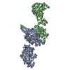
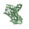
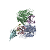
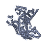
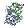
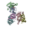
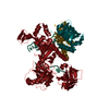
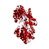
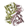
Entry Database : PDB / ID : 6iyoTitle Crystal Structure of the acyltransferase domain from the second module of the salinomycin polyketide synthase Type I modular polyketide synthase Keywords / / / Function / homology Function Domain/homology Component
/ / / / / / / / / / / / / / / / / / / / / / / / / / / / / / / / / / / / / / / / / / / / / / / / / / / / / / / / / / / / / / / / Biological species Streptomyces albus subsp. albus (bacteria)Method / / / Resolution : 2.2 Å Authors Zhang, F. / Zheng, J. Funding support Organization Grant number Country National Science Foundation (China) 31570056, 31770068
Journal : Biochemistry / Year : 2019Title : Structural Insights into the Substrate Specificity of Acyltransferases from Salinomycin Polyketide Synthase.Authors : Zhang, F. / Shi, T. / Ji, H. / Ali, I. / Huang, S. / Deng, Z. / Min, Q. / Bai, L. / Zhao, Y. / Zheng, J. History Deposition Dec 17, 2018 Deposition site / Processing site Revision 1.0 Jul 10, 2019 Provider / Type Revision 1.1 Jul 24, 2019 Group / Database references / Category / citation_authorItem _citation.journal_volume / _citation.page_first ... _citation.journal_volume / _citation.page_first / _citation.page_last / _citation_author.identifier_ORCID Revision 1.2 Nov 22, 2023 Group / Database references / Refinement descriptionCategory chem_comp_atom / chem_comp_bond ... chem_comp_atom / chem_comp_bond / database_2 / pdbx_initial_refinement_model Item / _database_2.pdbx_database_accession
Show all Show less
 Movie
Movie Controller

 Yorodumi
 Open data
 Basic information
 Components
 Keywords
 Function and homology information
 Streptomyces albus subsp. albus (bacteria)
 X-RAY DIFFRACTION /
  Authors
 China, 1items
  Citation
 Structure visualization
 Downloads & links
 Other downloads


 PDBj
PDBj


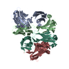
 Assembly
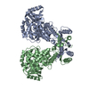
 Mass: 18.015 Da / Num. of mol.: 36 / Source method: isolated from a natural source / Formula: H2O
 Sample preparation
 Processing