 Movie
Movie Controller

[English] 日本語
 Yorodumi
- PDB-2abz: Crystal structure of C19A/C43A mutant of leech carboxypeptidase i... -
+ Open data

- Basic information
| Entry | Database: PDB / ID: 2abz | ||||||
|---|---|---|---|---|---|---|---|
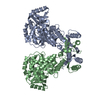
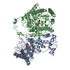
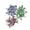
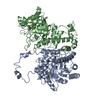
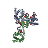
| Title | Crystal structure of C19A/C43A mutant of leech carboxypeptidase inhibitor in complex with bovine carboxypeptidase A | ||||||
 Components Components |
| ||||||
 Keywords Keywords | hydrolase/hydrolase inhibitor / inhibitor-metallocarboxypeptidase complex / LCI mutant / oxidative folding intermediate analog / hydrolase-hydrolase inhibitor COMPLEX | ||||||
| Function / homology |  Function and homology information Function and homology informationcarboxypeptidase A / leukotriene metabolic process / peptidase inhibitor activity / metallocarboxypeptidase activity / proteolysis / : / zinc ion binding Similarity search - Function | ||||||
| Biological species |   Hirudo medicinalis (medicinal leech) Hirudo medicinalis (medicinal leech) | ||||||
| Method |  X-RAY DIFFRACTION / MOLECULAR REPLACEMENT / Resolution: 2.16 Å X-RAY DIFFRACTION / MOLECULAR REPLACEMENT / Resolution: 2.16 Å | ||||||
 Authors Authors | Arolas, J.L. / Popowicz, G.M. / Bronsoms, S. / Aviles, F.X. / Huber, R. / Holak, T.A. / Ventura, S. | ||||||
 Citation Citation | Journal: J.Mol.Biol. / Year: 2005 Title: Study of a major intermediate in the oxidative folding of leech carboxypeptidase inhibitor: contribution of the fourth disulfide bond Authors: Arolas, J.L. / Popowicz, G.M. / Bronsoms, S. / Aviles, F.X. / Huber, R. / Holak, T.A. / Ventura, S. #1: Journal: NAT.STRUCT.MOL.BIOL. / Year: 2000Title: Structure of a novel leech carboxypeptidase inhibitor determined free in solution and in complex with human carboxypeptidase A2 Authors: Reverter, D. / Fernandez-Catalan, C. / Baumgartner, R. / Pfander, R. / Huber, R. / Bode, W. / Vendrell, J. / Holak, T.A. / Aviles, F.X. #2: Journal: Structure / Year: 2005Title: NMR structural characterization and computational predictions of the major intermediate in oxidative folding of leech carboxypeptidase inhibitor Authors: Arolas, J.L. / D'Silva, L. / Popowicz, G.M. / Aviles, F.X. / Holak, T.A. / Ventura, S. | ||||||
| History |
|
- Structure visualization
| Structure viewer | Molecule: MolmilJmol/JSmol |
|---|
- Downloads & links
-Download
| PDBx/mmCIF format | 2abz.cif.gz | 166 KB | Display | PDBx/mmCIF format |
|---|---|---|---|---|
| PDB format | pdb2abz.ent.gz | 133.2 KB | Display | PDB format |
| PDBx/mmJSON format | 2abz.json.gz | Tree view | PDBx/mmJSON format | |
| Others |  Other downloads Other downloads |
-Validation report
| Arichive directory | https://data.pdbj.org/pub/pdb/validation_reports/ab/2abzftp://data.pdbj.org/pub/pdb/validation_reports/ab/2abz | HTTPS FTP |
|---|
-Related structure data
| Related structure data | |
|---|---|
| Similar structure data |


-Links
 PDBj
PDBj
- Assembly
| Deposited unit | 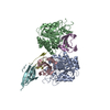
| ||||||||
|---|---|---|---|---|---|---|---|---|---|
| 1 |
| ||||||||
| Unit cell |
|
-Components
| #1: Protein | Mass: 34721.750 Da / Num. of mol.: 2 Source method: isolated from a genetically manipulated source Source: (gene. exp.)  Pichia pastoris (fungus) / Strain (production host): KM71 / References: UniProt: P00730, carboxypeptidase A Pichia pastoris (fungus) / Strain (production host): KM71 / References: UniProt: P00730, carboxypeptidase A#2: Protein | Mass: 7333.126 Da / Num. of mol.: 4 / Mutation: C19A/C43A Source method: isolated from a genetically manipulated source Source: (gene. exp.) Hirudo medicinalis (medicinal leech) / Plasmid: pBAT4 / Species (production host): Escherichia coli / Production host:  #3: Chemical |   Mass: 65.409 Da / Num. of mol.: 2 / Source method: obtained synthetically / Formula: Zn Mass: 65.409 Da / Num. of mol.: 2 / Source method: obtained synthetically / Formula: ZnHas protein modification | Y | |
|---|
-Experimental details
-Experiment
| Experiment | Method: X-RAY DIFFRACTION |
|---|
- Sample preparation
| Crystal | Density Matthews: 3.1 Å3/Da / Density % sol: 60.02 % |
|---|---|
| Crystal grow | Temperature: 293 K / Method: vapor diffusion, sitting drop / pH: 8.5 Details: 1.5M Lithium Sulfate monohydrate, 100mM Tris, pH 8.5, VAPOR DIFFUSION, SITTING DROP, temperature 293K |
-Data collection
| Detector | Date: Jun 21, 2004 |
|---|---|
| Radiation | Protocol: SINGLE WAVELENGTH / Monochromatic (M) / Laue (L): M / Scattering type: x-ray |
| Radiation wavelength | Relative weight: 1 |
| Reflection | Resolution: 2.1→30 Å / Num. obs: 66322 |
- Processing
| Software | Name: REFMAC / Version: 5.1.2 / Classification: refinement | ||||||||||||||||||||
|---|---|---|---|---|---|---|---|---|---|---|---|---|---|---|---|---|---|---|---|---|---|
| Refinement | Method to determine structure: MOLECULAR REPLACEMENT / Resolution: 2.16→30 Å / σ(F): 2 / Stereochemistry target values: Engh & Huber
| ||||||||||||||||||||
| Refinement step | Cycle: LAST / Resolution: 2.16→30 Å
|
