 Movie
Movie Controller

[English] 日本語
 Yorodumi
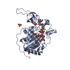
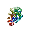
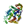
- PDB-1dtd: CRYSTAL STRUCTURE OF THE COMPLEX BETWEEN THE LEECH CARBOXYPEPTIDA... -
+ Open data

- Basic information
| Entry | Database: PDB / ID: 1dtd | ||||||
|---|---|---|---|---|---|---|---|
| Title | CRYSTAL STRUCTURE OF THE COMPLEX BETWEEN THE LEECH CARBOXYPEPTIDASE INHIBITOR AND THE HUMAN CARBOXYPEPTIDASE A2 (LCI-CPA2) | ||||||
 Components Components |
| ||||||
 Keywords Keywords | HYDROLASE/HYDROLASE INHIBITOR / Carboxypeptidase A2 / Leech Carboxypeptidase Inhibitor / HYDROLASE-HYDROLASE INHIBITOR COMPLEX | ||||||
| Function / homology |  Function and homology information Function and homology informationcarboxypeptidase A2 / protein catabolic process in the vacuole / Developmental Lineage of Pancreatic Acinar Cells / peptidase inhibitor activity / vacuole / carboxypeptidase activity / metallocarboxypeptidase activity / proteolysis / : / extracellular region / zinc ion binding Similarity search - Function | ||||||
| Biological species |  Homo sapiens (human) Homo sapiens (human) Hirudo medicinalis (medicinal leech) Hirudo medicinalis (medicinal leech) | ||||||
| Method |  X-RAY DIFFRACTION / Resolution: 1.65 Å X-RAY DIFFRACTION / Resolution: 1.65 Å | ||||||
 Authors Authors | Reverter, D. / Fernandez-Catalan, C. / Bode, W. / Holak, T.A. / Aviles, F.X. | ||||||
 Citation Citation | Journal: Nat.Struct.Biol. / Year: 2000 Title: Structure of a novel leech carboxypeptidase inhibitor determined free in solution and in complex with human carboxypeptidase A2. Authors: Reverter, D. / Fernandez-Catalan, C. / Baumgartner, R. / Pfander, R. / Huber, R. / Bode, W. / Vendrell, J. / Holak, T.A. / Aviles, F.X. | ||||||
| History |
|
- Structure visualization
| Structure viewer | Molecule: MolmilJmol/JSmol |
|---|
- Downloads & links
-Download
| PDBx/mmCIF format | 1dtd.cif.gz | 90.3 KB | Display | PDBx/mmCIF format |
|---|---|---|---|---|
| PDB format | pdb1dtd.ent.gz | 67.6 KB | Display | PDB format |
| PDBx/mmJSON format | 1dtd.json.gz | Tree view | PDBx/mmJSON format | |
| Others |  Other downloads Other downloads |
-Validation report
| Arichive directory | https://data.pdbj.org/pub/pdb/validation_reports/dt/1dtdftp://data.pdbj.org/pub/pdb/validation_reports/dt/1dtd | HTTPS FTP |
|---|
-Related structure data


-Links
 PDBj
PDBj


- Assembly
| Deposited unit | 
| ||||||||
|---|---|---|---|---|---|---|---|---|---|
| 1 |
| ||||||||
| Unit cell |
|
-Components
| #1: Protein | Mass: 33660.945 Da / Num. of mol.: 1 / Source method: isolated from a natural source / Source: (natural) Homo sapiens (human) / References: UniProt: P48052, peptidyl-dipeptidase A |
|---|---|
| #2: Protein | Mass: 6787.647 Da / Num. of mol.: 1 / Source method: isolated from a natural source / Source: (natural) Hirudo medicinalis (medicinal leech) / References: UniProt: P81511 |
| #3: Chemical | ChemComp-ZN /   Mass: 65.409 Da / Num. of mol.: 1 / Source method: obtained synthetically / Formula: Zn Mass: 65.409 Da / Num. of mol.: 1 / Source method: obtained synthetically / Formula: Zn |
| #4: Chemical | ChemComp-GLU /   Type: L-peptide linking / Mass: 147.129 Da / Num. of mol.: 1 / Source method: obtained synthetically / Formula: C5H9NO4 Type: L-peptide linking / Mass: 147.129 Da / Num. of mol.: 1 / Source method: obtained synthetically / Formula: C5H9NO4 |
| #5: Water | ChemComp-HOH /  Mass: 18.015 Da / Num. of mol.: 231 / Source method: isolated from a natural source / Formula: H2O Mass: 18.015 Da / Num. of mol.: 231 / Source method: isolated from a natural source / Formula: H2O |
| Has protein modification | Y |
-Experimental details
-Experiment
| Experiment | Method: X-RAY DIFFRACTION / Number of used crystals: 1 |
|---|
- Sample preparation
| Crystal | Density Matthews: 2.64 Å3/Da / Density % sol: 53.43 % | ||||||||||||||||||||||||||||||||||||
|---|---|---|---|---|---|---|---|---|---|---|---|---|---|---|---|---|---|---|---|---|---|---|---|---|---|---|---|---|---|---|---|---|---|---|---|---|---|
| Crystal grow | Temperature: 298 K / Method: vapor diffusion / pH: 7.5 Details: ammonium phosphate, sodium citrate, pH 7.5, VAPOR DIFFUSION, temperature 298.0K | ||||||||||||||||||||||||||||||||||||
| Crystal grow | *PLUS Temperature: 4, 20 ℃ / Method: vapor diffusion, sitting dropDetails: drop consists of equal volume of protein and reservoir solutions PH range low: 8.5 / PH range high: 6 | ||||||||||||||||||||||||||||||||||||
| Components of the solutions | *PLUS
|
-Data collection
| Diffraction | Mean temperature: 298 K |
|---|---|
| Diffraction source | Source: ROTATING ANODE / Type: RIGAKU / Wavelength: 1.54 |
| Detector | Type: RIGAKU / Detector: IMAGE PLATE / Date: Jan 1, 1999 |
| Radiation | Protocol: SINGLE WAVELENGTH / Monochromatic (M) / Laue (L): M / Scattering type: x-ray |
| Radiation wavelength | Wavelength: 1.54 Å / Relative weight: 1 |
| Reflection | Resolution: 1.65→20 Å / Num. all: 433570 / Num. obs: 52605 / % possible obs: 98 % / Observed criterion σ(F): 2 / Observed criterion σ(I): 5 / Redundancy: 5 % / Biso Wilson estimate: 10 Å2 / Rmerge(I) obs: 0.092 / Net I/σ(I): 80 |
| Reflection shell | Resolution: 1.65→1.75 Å / Redundancy: 6 % / Rmerge(I) obs: 0.15 / Num. unique all: 3567 / % possible all: 98 |
| Reflection | *PLUS Num. measured all: 433570 |
| Reflection shell | *PLUS % possible obs: 98 % |
- Processing
| Software |
| ||||||||||||||||||||
|---|---|---|---|---|---|---|---|---|---|---|---|---|---|---|---|---|---|---|---|---|---|
| Refinement | Resolution: 1.65→12 Å / σ(F): 2 / σ(I): 5
| ||||||||||||||||||||
| Refinement step | Cycle: LAST / Resolution: 1.65→12 Å
| ||||||||||||||||||||
| Refine LS restraints |
| ||||||||||||||||||||
| Software | *PLUS Name: REFMAC / Classification: refinement | ||||||||||||||||||||
| Refine LS restraints | *PLUS
|
