 Movie
Movie Controller

[English] 日本語
 Yorodumi
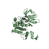
- PDB-6isu: Crystal structure of Lys27-linked di-ubiquitin in complex with it... -
+ Open data

- Basic information
| Entry | Database: PDB / ID: 6isu | ||||||||||||
|---|---|---|---|---|---|---|---|---|---|---|---|---|---|
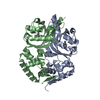
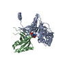
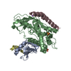
| Title | Crystal structure of Lys27-linked di-ubiquitin in complex with its selective interacting protein UCHL3 | ||||||||||||
 Components Components |
| ||||||||||||
 Keywords Keywords | HYDROLASE / Ubiquitination / UCHL3 / Lys27-linked di-ubiquitin | ||||||||||||
| Function / homology |  Function and homology information Function and homology informationdeNEDDylase activity / symbiont entry into host cell via disruption of host cell glycocalyx / symbiont entry into host cell via disruption of host cell envelope / virus tail / protein deubiquitination / Synthesis of active ubiquitin: roles of E1 and E2 enzymes / post-translational protein modification / ubiquitin binding / protein catabolic process / UCH proteinases ...deNEDDylase activity / symbiont entry into host cell via disruption of host cell glycocalyx / symbiont entry into host cell via disruption of host cell envelope / virus tail / protein deubiquitination / Synthesis of active ubiquitin: roles of E1 and E2 enzymes / post-translational protein modification / ubiquitin binding / protein catabolic process / UCH proteinases / Neddylation / peptidase activity / ubiquitin-dependent protein catabolic process / ubiquitinyl hydrolase 1 / cysteine-type deubiquitinase activity / protein ubiquitination / Golgi apparatus / nucleoplasm / cytosol / cytoplasm Similarity search - Function | ||||||||||||
| Biological species |  Homo sapiens (human) Homo sapiens (human) | ||||||||||||
| Method |  X-RAY DIFFRACTION / SYNCHROTRON / MOLECULAR REPLACEMENT / Resolution: 1.866 Å X-RAY DIFFRACTION / SYNCHROTRON / MOLECULAR REPLACEMENT / Resolution: 1.866 Å | ||||||||||||
 Authors Authors | Ding, S. / Pan, M. / Zheng, Q. / Ren, Y. / Hong, D. | ||||||||||||
| Funding support |  China, 3items China, 3items
| ||||||||||||
 Citation Citation | Journal: Angew. Chem. Int. Ed. Engl. / Year: 2019 Title: Chemical Protein Synthesis Enabled Mechanistic Studies on the Molecular Recognition of K27-linked Ubiquitin Chains. Authors: Pan, M. / Zheng, Q. / Ding, S. / Zhang, L. / Qu, Q. / Wang, T. / Hong, D. / Ren, Y. / Liang, L. / Chen, C. / Mei, Z. / Liu, L. | ||||||||||||
| History |
|
- Structure visualization
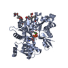
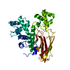
| Structure viewer | Molecule: MolmilJmol/JSmol |
|---|
- Downloads & links
-Download
| PDBx/mmCIF format | 6isu.cif.gz | 95.7 KB | Display | PDBx/mmCIF format |
|---|---|---|---|---|
| PDB format | pdb6isu.ent.gz | 71.3 KB | Display | PDB format |
| PDBx/mmJSON format | 6isu.json.gz | Tree view | PDBx/mmJSON format | |
| Others |  Other downloads Other downloads |
-Validation report
| Summary document | 6isu_validation.pdf.gz | 443.6 KB | Display | wwPDB validaton report |
|---|---|---|---|---|
| Full document | 6isu_full_validation.pdf.gz | 447.4 KB | Display | |
| Data in XML | 6isu_validation.xml.gz | 19.3 KB | Display | |
| Data in CIF | 6isu_validation.cif.gz | 27.9 KB | Display | |
| Arichive directory | https://data.pdbj.org/pub/pdb/validation_reports/is/6isuftp://data.pdbj.org/pub/pdb/validation_reports/is/6isu | HTTPS FTP |
-Related structure data
| Related structure data | 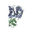 1xd3S S: Starting model for refinement |
|---|---|
| Similar structure data |


-Links
 PDBj
PDBj

- Assembly
| Deposited unit | 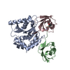
| |||||||||
|---|---|---|---|---|---|---|---|---|---|---|
| 1 |
| |||||||||
| Unit cell |
| |||||||||
| Components on special symmetry positions |
|
-Components
| #1: Protein | Mass: 26213.576 Da / Num. of mol.: 1 Source method: isolated from a genetically manipulated source Source: (gene. exp.) Homo sapiens (human) / Gene: UCHL3 / Production host:  |
|---|---|
| #2: Protein | Mass: 8576.831 Da / Num. of mol.: 1 Source method: isolated from a genetically manipulated source Source: (gene. exp.) Homo sapiens (human) / Gene: UBB / Production host: |
| #3: Protein | Mass: 8593.862 Da / Num. of mol.: 1 Source method: isolated from a genetically manipulated source Source: (gene. exp.) Homo sapiens (human) / Gene: UBB / Production host: |
| #4: Water | ChemComp-HOH /  Mass: 18.015 Da / Num. of mol.: 292 / Source method: isolated from a natural source / Formula: H2O Mass: 18.015 Da / Num. of mol.: 292 / Source method: isolated from a natural source / Formula: H2O |
| Has protein modification | Y |
-Experimental details
-Experiment
| Experiment | Method: X-RAY DIFFRACTION / Number of used crystals: 1 |
|---|
- Sample preparation
| Crystal | Density Matthews: 2.27 Å3/Da / Density % sol: 45.72 % |
|---|---|
| Crystal grow | Temperature: 291.15 K / Method: vapor diffusion, hanging drop / Details: 18% PEG 3350 (w/v), 400 mM Ca(AC)2 |
-Data collection
| Diffraction | Mean temperature: 77 K / Serial crystal experiment: N |
|---|---|
| Diffraction source | Source: SYNCHROTRON / Site: SSRF / Beamline: BL17B1 / Wavelength: 0.9792 Å |
| Detector | Type: BRUKER SMART 6000 / Detector: CCD / Date: Jan 10, 2018 |
| Radiation | Protocol: SINGLE WAVELENGTH / Monochromatic (M) / Laue (L): M / Scattering type: x-ray |
| Radiation wavelength | Wavelength: 0.9792 Å / Relative weight: 1 |
| Reflection | Resolution: 1.866→41.85 Å / Num. obs: 29404 / % possible obs: 88.82 % / Redundancy: 3.2 % / Net I/σ(I): 31.32 |
| Reflection shell | Resolution: 1.866→1.933 Å |
- Processing
| Software |
| |||||||||||||||||||||||||||||||||||||||||||||||||||||||||||||||||||||||||||||
|---|---|---|---|---|---|---|---|---|---|---|---|---|---|---|---|---|---|---|---|---|---|---|---|---|---|---|---|---|---|---|---|---|---|---|---|---|---|---|---|---|---|---|---|---|---|---|---|---|---|---|---|---|---|---|---|---|---|---|---|---|---|---|---|---|---|---|---|---|---|---|---|---|---|---|---|---|---|---|
| Refinement | Method to determine structure: MOLECULAR REPLACEMENT Starting model: 1XD3 Resolution: 1.866→41.85 Å / SU ML: 0.25 / Cross valid method: FREE R-VALUE / Phase error: 33.09
| |||||||||||||||||||||||||||||||||||||||||||||||||||||||||||||||||||||||||||||
| Solvent computation | Shrinkage radii: 0.9 Å / VDW probe radii: 1.11 Å | |||||||||||||||||||||||||||||||||||||||||||||||||||||||||||||||||||||||||||||
| Refinement step | Cycle: LAST / Resolution: 1.866→41.85 Å
| |||||||||||||||||||||||||||||||||||||||||||||||||||||||||||||||||||||||||||||
| Refine LS restraints |
| |||||||||||||||||||||||||||||||||||||||||||||||||||||||||||||||||||||||||||||
| LS refinement shell |
|
