 Movie
Movie Controller

[English] 日本語
 Yorodumi
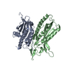
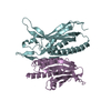
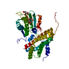
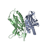
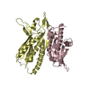
- PDB-4f48: The X-ray structural of FimXEAL-c-di-GMP-PilZ complexes from Xant... -
+ Open data

- Basic information
| Entry | Database: PDB / ID: 4f48 | ||||||
|---|---|---|---|---|---|---|---|
| Title | The X-ray structural of FimXEAL-c-di-GMP-PilZ complexes from Xanthomonas campestris | ||||||
 Components Components |
| ||||||
 Keywords Keywords | SIGNALING PROTEIN / FimXEAL-c-di-GMP-PilZ complex / type IV pilus | ||||||
| Function / homology |  Function and homology information Function and homology informationregulation of single-species biofilm formation / cyclic-guanylate-specific phosphodiesterase activity / cyclic-di-GMP binding / nucleotide binding / plasma membrane Similarity search - Function | ||||||
| Biological species |  Xanthomonas campestris pv. campestris (bacteria) Xanthomonas campestris pv. campestris (bacteria) | ||||||
| Method |  X-RAY DIFFRACTION / SYNCHROTRON / SAD / Resolution: 3 Å X-RAY DIFFRACTION / SYNCHROTRON / SAD / Resolution: 3 Å | ||||||
 Authors Authors | Chin, K.-H. / Liao, Y.-T. / Chou, S.-H. | ||||||
 Citation Citation | Journal: Acta Crystallogr.,Sect.D / Year: 2012 Title: Structural polymorphism of c-di-GMP bound to an EAL domain and in complex with a type II PilZ-domain protein. Authors: Chin, K.-H. / Kuo, W.-T. / Yu, Y.-J. / Liao, Y.-T. / Yang, M.T. / Chou, S.-H. | ||||||
| History |
|
- Structure visualization
| Structure viewer | Molecule: MolmilJmol/JSmol |
|---|
- Downloads & links
-Download
| PDBx/mmCIF format | 4f48.cif.gz | 82.9 KB | Display | PDBx/mmCIF format |
|---|---|---|---|---|
| PDB format | pdb4f48.ent.gz | 61.9 KB | Display | PDB format |
| PDBx/mmJSON format | 4f48.json.gz | Tree view | PDBx/mmJSON format | |
| Others |  Other downloads Other downloads |
-Validation report
| Arichive directory | https://data.pdbj.org/pub/pdb/validation_reports/f4/4f48ftp://data.pdbj.org/pub/pdb/validation_reports/f4/4f48 | HTTPS FTP |
|---|
-Related structure data


-Links
 PDBj
PDBj


- Assembly
| Deposited unit | 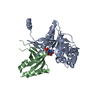
| ||||||||
|---|---|---|---|---|---|---|---|---|---|
| 1 |
| ||||||||
| 2 | 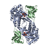
| ||||||||
| Unit cell |
| ||||||||
| Components on special symmetry positions |
|
-Components
| #1: Protein | Mass: 28074.807 Da / Num. of mol.: 1 / Fragment: UNP residues 437-684 Source method: isolated from a genetically manipulated source Source: (gene. exp.) Xanthomonas campestris pv. campestris (bacteria)Gene: XCC2291 / Production host: |
|---|---|
| #2: Protein | Mass: 12433.372 Da / Num. of mol.: 1 Source method: isolated from a genetically manipulated source Source: (gene. exp.) Xanthomonas campestris pv. campestris (bacteria)Gene: pilZ, XCC1024 / Production host: |
| #3: Chemical | ChemComp-C2E / 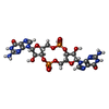  Mass: 690.411 Da / Num. of mol.: 1 / Source method: obtained synthetically / Formula: C20H24N10O14P2 Mass: 690.411 Da / Num. of mol.: 1 / Source method: obtained synthetically / Formula: C20H24N10O14P2 |
| #4: Water | ChemComp-HOH /  Mass: 18.015 Da / Num. of mol.: 80 / Source method: isolated from a natural source / Formula: H2O Mass: 18.015 Da / Num. of mol.: 80 / Source method: isolated from a natural source / Formula: H2O |
-Experimental details
-Experiment
| Experiment | Method: X-RAY DIFFRACTION / Number of used crystals: 6 |
|---|
- Sample preparation
| Crystal | Density Matthews: 2.89 Å3/Da / Density % sol: 57.45 % |
|---|---|
| Crystal grow | Temperature: 298 K / Method: vapor diffusion, sitting drop / pH: 8 Details: 0.1M HEPES pH 7.5, 20% PEG 3000, VAPOR DIFFUSION, SITTING DROP, temperature 298K |
-Data collection
| Diffraction | Mean temperature: 100 K |
|---|---|
| Diffraction source | Source: SYNCHROTRON / Site: NSRRC  / Beamline: BL13B1 / Wavelength: 0.97934 Å / Beamline: BL13B1 / Wavelength: 0.97934 Å |
| Detector | Type: ADSC QUANTUM 315 / Detector: CCD / Date: Apr 15, 2011 |
| Radiation | Monochromator: GRAPHITE / Protocol: SINGLE WAVELENGTH / Monochromatic (M) / Laue (L): M / Scattering type: x-ray |
| Radiation wavelength | Wavelength: 0.97934 Å / Relative weight: 1 |
| Reflection | Resolution: 2.7→30 Å / Num. obs: 27412 / % possible obs: 99.4 % / Observed criterion σ(F): 2 / Observed criterion σ(I): 2 / Biso Wilson estimate: 22.9 Å2 |
| Reflection shell | Highest resolution: 2.7 Å / % possible all: 99.4 |
- Processing
| Software |
| ||||||||||||||||||||||||||||||||||||||||||||||||||||||||||||||||||||||||||||||||
|---|---|---|---|---|---|---|---|---|---|---|---|---|---|---|---|---|---|---|---|---|---|---|---|---|---|---|---|---|---|---|---|---|---|---|---|---|---|---|---|---|---|---|---|---|---|---|---|---|---|---|---|---|---|---|---|---|---|---|---|---|---|---|---|---|---|---|---|---|---|---|---|---|---|---|---|---|---|---|---|---|---|
| Refinement | Method to determine structure: SAD / Resolution: 3→22.56 Å / Rfactor Rfree error: 0.01 / Data cutoff high absF: 69323.4 / Data cutoff low absF: 0 / Isotropic thermal model: RESTRAINED / Cross valid method: THROUGHOUT / σ(F): 0 / Stereochemistry target values: Engh & Huber Details: 1. BULK SOLVENT MODEL USED 2. FRIEDEL PAIRS WERE CONSIDERED SEPARATELY AS UNIQUE REFLECTIONS IN THE CALCULATION OF THE STATISTICS.
| ||||||||||||||||||||||||||||||||||||||||||||||||||||||||||||||||||||||||||||||||
| Solvent computation | Solvent model: FLAT MODEL / Bsol: 22.9565 Å2 / ksol: 0.35 e/Å3 | ||||||||||||||||||||||||||||||||||||||||||||||||||||||||||||||||||||||||||||||||
| Displacement parameters | Biso mean: 27.8 Å2
| ||||||||||||||||||||||||||||||||||||||||||||||||||||||||||||||||||||||||||||||||
| Refine analyze |
| ||||||||||||||||||||||||||||||||||||||||||||||||||||||||||||||||||||||||||||||||
| Refinement step | Cycle: LAST / Resolution: 3→22.56 Å
| ||||||||||||||||||||||||||||||||||||||||||||||||||||||||||||||||||||||||||||||||
| Refine LS restraints |
| ||||||||||||||||||||||||||||||||||||||||||||||||||||||||||||||||||||||||||||||||
| Refine LS restraints NCS | NCS model details: NONE | ||||||||||||||||||||||||||||||||||||||||||||||||||||||||||||||||||||||||||||||||
| LS refinement shell | Resolution: 2.8→2.98 Å / Rfactor Rfree error: 0.032 / Total num. of bins used: 6
| ||||||||||||||||||||||||||||||||||||||||||||||||||||||||||||||||||||||||||||||||
| Xplor file |
|
