 Movie
Movie Controller


+ Open data

- Basic information
| Entry | Database: PDB / ID: 6ioc | |||||||||
|---|---|---|---|---|---|---|---|---|---|---|
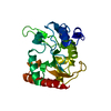
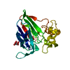
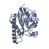
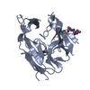
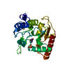
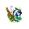
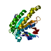
| Title | The structure of the H109Q mutant of UdgX in complex with uracil | |||||||||
 Components Components | Phage SPO1 DNA polymerase-related protein | |||||||||
 Keywords Keywords | HYDROLASE / uracil DNA glycosylase / DNA repair / iron-sulfur | |||||||||
| Function / homology |  Function and homology information Function and homology informationdeaminated base DNA N-glycosylase activity / nucleotidyltransferase activity / 4 iron, 4 sulfur cluster binding / DNA repair / metal ion binding Similarity search - Function | |||||||||
| Biological species |  Mycobacterium smegmatis MC2 155 (bacteria) Mycobacterium smegmatis MC2 155 (bacteria) | |||||||||
| Method |  X-RAY DIFFRACTION / SYNCHROTRON / MOLECULAR REPLACEMENT / Resolution: 1.624 Å X-RAY DIFFRACTION / SYNCHROTRON / MOLECULAR REPLACEMENT / Resolution: 1.624 Å | |||||||||
 Authors Authors | Xie, W. / Tu, J. | |||||||||
| Funding support |  China, 2items China, 2items
| |||||||||
 Citation Citation | Journal: Nat.Chem.Biol. / Year: 2019 Title: Suicide inactivation of the uracil DNA glycosylase UdgX by covalent complex formation. Authors: Tu, J. / Chen, R. / Yang, Y. / Cao, W. / Xie, W. | |||||||||
| History |
|
- Structure visualization
| Structure viewer | Molecule: MolmilJmol/JSmol |
|---|
- Downloads & links
-Download
| PDBx/mmCIF format | 6ioc.cif.gz | 58.8 KB | Display | PDBx/mmCIF format |
|---|---|---|---|---|
| PDB format | pdb6ioc.ent.gz | 39.6 KB | Display | PDB format |
| PDBx/mmJSON format | 6ioc.json.gz | Tree view | PDBx/mmJSON format | |
| Others |  Other downloads Other downloads |
-Validation report
| Arichive directory | https://data.pdbj.org/pub/pdb/validation_reports/io/6iocftp://data.pdbj.org/pub/pdb/validation_reports/io/6ioc | HTTPS FTP |
|---|
-Related structure data
| Related structure data |  6io9C  6ioaSC  6iobC  6iodC S: Starting model for refinement C: citing same article ( |
|---|---|
| Similar structure data |


-Links
 PDBj
PDBj- Assembly
| Deposited unit | 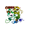
| ||||||||
|---|---|---|---|---|---|---|---|---|---|
| 1 |
| ||||||||
| Unit cell |
|
-Components
| #1: Protein | Mass: 22473.541 Da / Num. of mol.: 1 / Fragment: UNP residues 7-215 / Mutation: H109Q Source method: isolated from a genetically manipulated source Source: (gene. exp.) Mycobacterium smegmatis MC2 155 (bacteria)Strain: mc2 155 / Gene: MSMEI_0259 / Production host: |
|---|---|
| #2: Chemical | ChemComp-SF4 /   Mass: 351.640 Da / Num. of mol.: 1 / Source method: obtained synthetically / Formula: Fe4S4 / Feature type: SUBJECT OF INVESTIGATION Mass: 351.640 Da / Num. of mol.: 1 / Source method: obtained synthetically / Formula: Fe4S4 / Feature type: SUBJECT OF INVESTIGATION |
| #3: Chemical | ChemComp-URA /   Mass: 112.087 Da / Num. of mol.: 1 / Source method: obtained synthetically / Formula: C4H4N2O2 / Feature type: SUBJECT OF INVESTIGATION Mass: 112.087 Da / Num. of mol.: 1 / Source method: obtained synthetically / Formula: C4H4N2O2 / Feature type: SUBJECT OF INVESTIGATION |
| #4: Water | ChemComp-HOH /  Mass: 18.015 Da / Num. of mol.: 146 / Source method: isolated from a natural source / Formula: H2O Mass: 18.015 Da / Num. of mol.: 146 / Source method: isolated from a natural source / Formula: H2O |
-Experimental details
-Experiment
| Experiment | Method: X-RAY DIFFRACTION / Number of used crystals: 1 |
|---|
- Sample preparation
| Crystal | Density Matthews: 1.69 Å3/Da / Density % sol: 27.41 % |
|---|---|
| Crystal grow | Temperature: 298 K / Method: vapor diffusion, sitting drop / Details: 2.15M Sodium malonate pH 7.0 and 3.8% MPD / PH range: 7 |
-Data collection
| Diffraction | Mean temperature: 100 K / Serial crystal experiment: N |
|---|---|
| Diffraction source | Source: SYNCHROTRON / Site: NFPSS / Beamline: BL19U1 / Wavelength: 0.979 Å |
| Detector | Type: DECTRIS PILATUS3 S 6M / Detector: PIXEL / Date: Sep 23, 2018 |
| Radiation | Protocol: SINGLE WAVELENGTH / Monochromatic (M) / Laue (L): M / Scattering type: x-ray |
| Radiation wavelength | Wavelength: 0.979 Å / Relative weight: 1 |
| Reflection | Resolution: 1.62→50 Å / Num. obs: 18944 / % possible obs: 99.5 % / Observed criterion σ(I): 3.8 / Redundancy: 6 % / CC1/2: 0.973 / Rmerge(I) obs: 0.12 / Net I/σ(I): 14.9 |
| Reflection shell | Resolution: 1.62→1.65 Å / Redundancy: 4.7 % / Rmerge(I) obs: 0.57 / Mean I/σ(I) obs: 3.8 / Num. unique obs: 914 / CC1/2: 0.876 / % possible all: 99.9 |
- Processing
| Software |
| |||||||||||||||||||||||||||||||||||||||||||||||||
|---|---|---|---|---|---|---|---|---|---|---|---|---|---|---|---|---|---|---|---|---|---|---|---|---|---|---|---|---|---|---|---|---|---|---|---|---|---|---|---|---|---|---|---|---|---|---|---|---|---|---|
| Refinement | Method to determine structure: MOLECULAR REPLACEMENT Starting model: 6IOA Resolution: 1.624→30.827 Å / SU ML: 0.16 / Cross valid method: FREE R-VALUE / σ(F): 1.36 / Phase error: 19.35
| |||||||||||||||||||||||||||||||||||||||||||||||||
| Solvent computation | Shrinkage radii: 0.9 Å / VDW probe radii: 1.11 Å | |||||||||||||||||||||||||||||||||||||||||||||||||
| Refinement step | Cycle: LAST / Resolution: 1.624→30.827 Å
| |||||||||||||||||||||||||||||||||||||||||||||||||
| Refine LS restraints |
| |||||||||||||||||||||||||||||||||||||||||||||||||
| LS refinement shell |
|
