- PDB-6in4: Crystal structure of apo DAPK1 in the presence of 18-crown-6 -
+
Open data
ID or keywords:
Loading...
-
Basic information
Entry
Database: PDB / ID: 6in4
Title
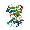
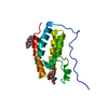
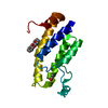
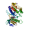
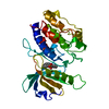
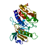
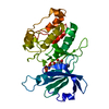
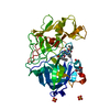
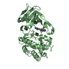
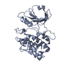
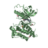
Crystal structure of apo DAPK1 in the presence of 18-crown-6
Components
Death-associated protein kinase 1
Keywords
TRANSFERASE / serine/threonine protein kinase / cell death
Function / homology
Function and homology information
cellular response to hydroperoxide / negative regulation of kinase activity / regulation of response to tumor cell / positive regulation of autophagic cell death / Caspase activation via Dependence Receptors in the absence of ligand / defense response to tumor cell / calcium/calmodulin-dependent protein kinase activity / regulation of NMDA receptor activity / syntaxin-1 binding / positive regulation of autophagy ...cellular response to hydroperoxide / negative regulation of kinase activity / regulation of response to tumor cell / positive regulation of autophagic cell death / Caspase activation via Dependence Receptors in the absence of ligand / defense response to tumor cell / calcium/calmodulin-dependent protein kinase activity / regulation of NMDA receptor activity / syntaxin-1 binding / positive regulation of autophagy / regulation of autophagy / apoptotic signaling pathway / cellular response to type II interferon / protein autophosphorylation / actin cytoskeleton / regulation of apoptotic process / protein phosphorylation / protein kinase activity / calmodulin binding / non-specific serine/threonine protein kinase / negative regulation of translation / postsynaptic density / intracellular signal transduction / positive regulation of apoptotic process / protein serine kinase activity / protein serine/threonine kinase activity / apoptotic process / negative regulation of apoptotic process / GTP binding / glutamatergic synapse / ATP binding / identical protein binding / nucleus / plasma membrane / cytoplasm Similarity search - Function
Death-associated protein kinase 1 / Roc domain profile. / Roc domain / Death domain profile. / DEATH domain, found in proteins involved in cell death (apoptosis). / Death domain / Death domain / Ankyrin repeats (many copies) / Death-like domain superfamily / Ankyrin repeat ...Death-associated protein kinase 1 / Roc domain profile. / Roc domain / Death domain profile. / DEATH domain, found in proteins involved in cell death (apoptosis). / Death domain / Death domain / Ankyrin repeats (many copies) / Death-like domain superfamily / Ankyrin repeat / Ankyrin repeat profile. / Ankyrin repeats (3 copies) / Ankyrin repeat region circular profile. / ankyrin repeats / Ankyrin repeat / Ankyrin repeat-containing domain superfamily / Phosphorylase Kinase; domain 1 / Phosphorylase Kinase; domain 1 / Transferase(Phosphotransferase) domain 1 / Transferase(Phosphotransferase); domain 1 / Serine/threonine-protein kinase, active site / Serine/Threonine protein kinases active-site signature. / Protein kinase domain / Serine/Threonine protein kinases, catalytic domain / Protein kinase, ATP binding site / Protein kinases ATP-binding region signature. / Protein kinase domain profile. / Protein kinase domain / Protein kinase-like domain superfamily / P-loop containing nucleoside triphosphate hydrolase / 2-Layer Sandwich / Orthogonal Bundle / Mainly Alpha / Alpha Beta Similarity search - Domain/homology
Mass: 33796.402 Da / Num. of mol.: 1 Source method: isolated from a genetically manipulated source Source: (gene. exp.) Homo sapiens (human) / Gene: DAPK1, DAPK / Production host: Escherichia coli (E. coli) References: UniProt: P53355, non-specific serine/threonine protein kinase
In the structure databanks used in Yorodumi, some data are registered as the other names, "COVID-19 virus" and "2019-nCoV". Here are the details of the virus and the list of structure data.
Jan 31, 2019. EMDB accession codes are about to change! (news from PDBe EMDB page)
EMDB accession codes are about to change! (news from PDBe EMDB page)
The allocation of 4 digits for EMDB accession codes will soon come to an end. Whilst these codes will remain in use, new EMDB accession codes will include an additional digit and will expand incrementally as the available range of codes is exhausted. The current 4-digit format prefixed with “EMD-” (i.e. EMD-XXXX) will advance to a 5-digit format (i.e. EMD-XXXXX), and so on. It is currently estimated that the 4-digit codes will be depleted around Spring 2019, at which point the 5-digit format will come into force.
The EM Navigator/Yorodumi systems omit the EMD- prefix.
Related info.:Q: What is EMD? / ID/Accession-code notation in Yorodumi/EM Navigator
Yorodumi is a browser for structure data from EMDB, PDB, SASBDB, etc.
This page is also the successor to EM Navigator detail page, and also detail information page/front-end page for Omokage search.
The word "yorodu" (or yorozu) is an old Japanese word meaning "ten thousand". "mi" (miru) is to see.
Related info.:EMDB / PDB / SASBDB / Comparison of 3 databanks / Yorodumi Search / Aug 31, 2016. New EM Navigator & Yorodumi / Yorodumi Papers / Jmol/JSmol / Function and homology information / Changes in new EM Navigator and Yorodumi
 Movie
Movie Controller


 Open data
 Basic information
 Components
 Keywords
 Function and homology information
 Homo sapiens (human)
 X-RAY DIFFRACTION /
  Authors
 Citation
 Structure visualization
 Downloads & links
 Other downloads
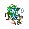


 PDBj
PDBj


 Assembly
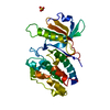


 Mass: 96.063 Da / Num. of mol.: 1 / Source method: obtained synthetically / Formula: SO4
 Mass: 18.015 Da / Num. of mol.: 235 / Source method: isolated from a natural source / Formula: H2O
 Sample preparation
 / Beamline: BL-5A / Wavelength: 1 Å
 Processing