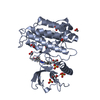
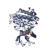
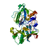
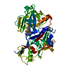
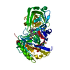
Entry Database : PDB / ID : 5a4cTitle FGFR1 ligand complex FIBROBLAST GROWTH FACTOR RECEPTOR 1 (FMS-RELATED TYROSINE KINASE 2, PFEIFFER SYNDROME), ISOFORM CRA_B Keywords / Function / homology Function Domain/homology Component
/ / / / / / / / / / / / / / / / / / / / / / / / / / / / / / / / / / / / / / / / / / / / / / / / / / / / / / / / / / / / / / / / / / / / / / / / / / / / / / / / / / / / / / / / / / / / / / / / / / / / / / / / / / / / / / / / / / / / / / / / / / / / / / / / / / / / / / / / / / / / / / / / / / / / / / Biological species HOMO SAPIENS (human)Method / / / Resolution : 2.09 Å Authors Klein, T. / Vajpai, N. / Phillips, J.J. / Davies, G. / Holdgate, G.A. / Phillips, C. / Tucker, J.A. / Norman, R.A. / Scott, A.S. / Higazi, D.R. ...Klein, T. / Vajpai, N. / Phillips, J.J. / Davies, G. / Holdgate, G.A. / Phillips, C. / Tucker, J.A. / Norman, R.A. / Scott, A.S. / Higazi, D.R. / Lowe, D. / Thompson, G.S. / Breeze, A.L. Journal : Nat.Commun. / Year : 2015Title : Structural and Dynamic Insights Into the Energetics of Activation Loop Rearrangement in Fgfr1 Kinase.Authors : Klein, T. / Vajpai, N. / Phillips, J.J. / Davies, G. / Holdgate, G.A. / Phillips, C. / Tucker, J.A. / Norman, R.A. / Scott, A.D. / Higazi, D.R. / Lowe, D. / Thompson, G.S. / Breeze, A.L. History Deposition Jun 5, 2015 Deposition site / Processing site Revision 1.0 Aug 5, 2015 Provider / Type Revision 1.1 May 8, 2024 Group Data collection / Database references ... Data collection / Database references / Derived calculations / Other Category chem_comp_atom / chem_comp_bond ... chem_comp_atom / chem_comp_bond / database_2 / pdbx_database_status / struct_site Item _database_2.pdbx_DOI / _database_2.pdbx_database_accession ... _database_2.pdbx_DOI / _database_2.pdbx_database_accession / _pdbx_database_status.status_code_sf / _struct_site.pdbx_auth_asym_id / _struct_site.pdbx_auth_comp_id / _struct_site.pdbx_auth_seq_id
Show all Show less
 Movie
Movie Controller


 Open data
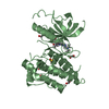
 Basic information
 Components
 Keywords
 Function and homology information
 HOMO SAPIENS (human)
 X-RAY DIFFRACTION /
  Authors
 Citation
 Structure visualization
 Downloads & links
 Other downloads
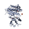

 PDBj
PDBj


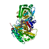
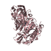
 Assembly
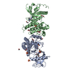


 Mass: 96.063 Da / Num. of mol.: 6 / Source method: obtained synthetically / Formula: SO4
 Mass: 62.068 Da / Num. of mol.: 10 / Source method: obtained synthetically / Formula: C2H6O2
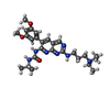
 Mass: 509.644 Da / Num. of mol.: 2 / Source method: obtained synthetically / Formula: C27H39N7O3
 Mass: 18.015 Da / Num. of mol.: 347 / Source method: isolated from a natural source / Formula: H2O
 Sample preparation
 / Beamline: I04 / Wavelength: 1.54
  Processing