 Movie
Movie Controller

[English] 日本語
 Yorodumi
- PDB-6im4: Structural basis of AimP signaling molecule recognition by AimR i... -
+ Open data

- Basic information
| Entry | Database: PDB / ID: 6im4 | ||||||
|---|---|---|---|---|---|---|---|
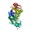
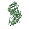
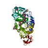
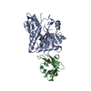
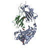
| Title | Structural basis of AimP signaling molecule recognition by AimR in Spbeta group of bacteriophages | ||||||
 Components Components |
| ||||||
 Keywords Keywords | PEPTIDE BINDING PROTEIN / Receptor / VIRUS | ||||||
| Function / homology | : / AimR transcriptional regulator-like / latency-replication decision / AimR transcriptional regulator Function and homology information Function and homology information | ||||||
| Biological species |  Bacillus phage SPbeta (virus) Bacillus phage SPbeta (virus)synthetic construct (others) | ||||||
| Method |  X-RAY DIFFRACTION / SYNCHROTRON / MOLECULAR REPLACEMENT / Resolution: 1.93 Å X-RAY DIFFRACTION / SYNCHROTRON / MOLECULAR REPLACEMENT / Resolution: 1.93 Å | ||||||
 Authors Authors | Ouyang, S.Y. | ||||||
 Citation Citation | Journal: Protein Cell / Year: 2019 Title: Structural basis of AimP signaling molecule recognition by AimR in Spbeta group of bacteriophages. Authors: Zhen, X. / Zhou, H. / Ding, W. / Zhou, B. / Xu, X. / Perculija, V. / Chen, C.J. / Chang, M.X. / Choudhary, M.I. / Ouyang, S. | ||||||
| History |
|
- Structure visualization
| Structure viewer | Molecule: MolmilJmol/JSmol |
|---|
- Downloads & links
-Download
| PDBx/mmCIF format | 6im4.cif.gz | 173.6 KB | Display | PDBx/mmCIF format |
|---|---|---|---|---|
| PDB format | pdb6im4.ent.gz | 139.7 KB | Display | PDB format |
| PDBx/mmJSON format | 6im4.json.gz | Tree view | PDBx/mmJSON format | |
| Others |  Other downloads Other downloads |
-Validation report
| Arichive directory | https://data.pdbj.org/pub/pdb/validation_reports/im/6im4ftp://data.pdbj.org/pub/pdb/validation_reports/im/6im4 | HTTPS FTP |
|---|
-Related structure data
| Related structure data |  6ipxC  6im2 C: citing same article ( |
|---|---|
| Similar structure data |


-Links
 PDBj
PDBj- Assembly
| Deposited unit | 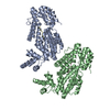
| ||||||||
|---|---|---|---|---|---|---|---|---|---|
| 1 | 
| ||||||||
| 2 | 
| ||||||||
| Unit cell |
|
-Components
| #1: Protein | Mass: 45849.477 Da / Num. of mol.: 2 Source method: isolated from a genetically manipulated source Source: (gene. exp.) Bacillus phage SPbeta (virus) / Gene: aimR, yopK / Production host:  #2: Protein/peptide | Mass: 588.701 Da / Num. of mol.: 2 / Source method: obtained synthetically / Source: (synth.) synthetic construct (others) #3: Water | ChemComp-HOH / |  Mass: 18.015 Da / Num. of mol.: 314 / Source method: isolated from a natural source / Formula: H2O Mass: 18.015 Da / Num. of mol.: 314 / Source method: isolated from a natural source / Formula: H2O |
|---|
-Experimental details
-Experiment
| Experiment | Method: X-RAY DIFFRACTION / Number of used crystals: 1 |
|---|
- Sample preparation
| Crystal | Density Matthews: 2.36 Å3/Da / Density % sol: 47.87 % |
|---|---|
| Crystal grow | Temperature: 291.5 K / Method: vapor diffusion, sitting drop / pH: 8.5 / Details: PEG 4000 magnesium acetate |
-Data collection
| Diffraction | Mean temperature: 100 K / Serial crystal experiment: N |
|---|---|
| Diffraction source | Source: SYNCHROTRON / Site: SSRF  / Beamline: BL17U1 / Wavelength: 0.979183 Å / Beamline: BL17U1 / Wavelength: 0.979183 Å |
| Detector | Type: ADSC QUANTUM 315r / Detector: CCD / Date: Dec 18, 2017 |
| Radiation | Protocol: SINGLE WAVELENGTH / Monochromatic (M) / Laue (L): M / Scattering type: x-ray |
| Radiation wavelength | Wavelength: 0.979183 Å / Relative weight: 1 |
| Reflection | Resolution: 1.928→61.565 Å / Num. obs: 135748 / % possible obs: 96.73 % / Redundancy: 10 % / CC1/2: 0.999 / Rmerge(I) obs: 0.106 / Rsym value: 0.111 / Net I/σ(I): 2 |
| Reflection shell | Resolution: 1.93→2.03 Å |
- Processing
| Software |
| ||||||||||||||||||||||||||||||||||||||||||||||||||||||||||||||||||||||||||||||||||||||||||||||||||||||||||||||||||||||||||||||||||||||||||||||||||||||||||||||||||||||||||||||||||||||||||||||||||||
|---|---|---|---|---|---|---|---|---|---|---|---|---|---|---|---|---|---|---|---|---|---|---|---|---|---|---|---|---|---|---|---|---|---|---|---|---|---|---|---|---|---|---|---|---|---|---|---|---|---|---|---|---|---|---|---|---|---|---|---|---|---|---|---|---|---|---|---|---|---|---|---|---|---|---|---|---|---|---|---|---|---|---|---|---|---|---|---|---|---|---|---|---|---|---|---|---|---|---|---|---|---|---|---|---|---|---|---|---|---|---|---|---|---|---|---|---|---|---|---|---|---|---|---|---|---|---|---|---|---|---|---|---|---|---|---|---|---|---|---|---|---|---|---|---|---|---|---|---|---|---|---|---|---|---|---|---|---|---|---|---|---|---|---|---|---|---|---|---|---|---|---|---|---|---|---|---|---|---|---|---|---|---|---|---|---|---|---|---|---|---|---|---|---|---|---|---|---|
| Refinement | Method to determine structure: MOLECULAR REPLACEMENT / Resolution: 1.93→61.565 Å / SU ML: 0.3 / Cross valid method: NONE / σ(F): 1.35 / Phase error: 32.32
| ||||||||||||||||||||||||||||||||||||||||||||||||||||||||||||||||||||||||||||||||||||||||||||||||||||||||||||||||||||||||||||||||||||||||||||||||||||||||||||||||||||||||||||||||||||||||||||||||||||
| Solvent computation | Shrinkage radii: 0.9 Å / VDW probe radii: 1.11 Å | ||||||||||||||||||||||||||||||||||||||||||||||||||||||||||||||||||||||||||||||||||||||||||||||||||||||||||||||||||||||||||||||||||||||||||||||||||||||||||||||||||||||||||||||||||||||||||||||||||||
| Refinement step | Cycle: LAST / Resolution: 1.93→61.565 Å
| ||||||||||||||||||||||||||||||||||||||||||||||||||||||||||||||||||||||||||||||||||||||||||||||||||||||||||||||||||||||||||||||||||||||||||||||||||||||||||||||||||||||||||||||||||||||||||||||||||||
| Refine LS restraints |
| ||||||||||||||||||||||||||||||||||||||||||||||||||||||||||||||||||||||||||||||||||||||||||||||||||||||||||||||||||||||||||||||||||||||||||||||||||||||||||||||||||||||||||||||||||||||||||||||||||||
| LS refinement shell |
|
