 Movie
Movie Controller

[English] 日本語
 Yorodumi
- PDB-6tp0: Crystal structure of Bacillus paralicheniformis alpha-amylase in ... -
+ Open data

- Basic information
| Entry | Database: PDB / ID: 6tp0 | ||||||
|---|---|---|---|---|---|---|---|
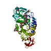
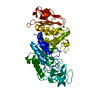
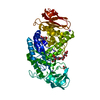
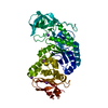
| Title | Crystal structure of Bacillus paralicheniformis alpha-amylase in complex with maltose | ||||||
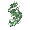
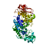
 Components Components | Amylase | ||||||
 Keywords Keywords | HYDROLASE / amylase / starch binding site | ||||||
| Function / homology |  Function and homology information Function and homology informationalpha-amylase / alpha-amylase activity / carbohydrate metabolic process / calcium ion binding / extracellular region Similarity search - Function | ||||||
| Biological species |  | ||||||
| Method |  X-RAY DIFFRACTION / FOURIER SYNTHESIS / Resolution: 2.04 Å X-RAY DIFFRACTION / FOURIER SYNTHESIS / Resolution: 2.04 Å | ||||||
 Authors Authors | Rozeboom, H.J. / Janssen, D.B. | ||||||
 Citation Citation | Journal: Int.J.Biol.Macromol. / Year: 2020 Title: Characterization of the starch surface binding site on Bacillus paralicheniformis alpha-amylase. Authors: Bozic, N. / Rozeboom, H.J. / Loncar, N. / Slavic, M.S. / Janssen, D.B. / Vujcic, Z. | ||||||
| History |
|
- Structure visualization
| Structure viewer | Molecule: MolmilJmol/JSmol |
|---|
- Downloads & links
-Download
| PDBx/mmCIF format | 6tp0.cif.gz | 213.3 KB | Display | PDBx/mmCIF format |
|---|---|---|---|---|
| PDB format | pdb6tp0.ent.gz | 168.8 KB | Display | PDB format |
| PDBx/mmJSON format | 6tp0.json.gz | Tree view | PDBx/mmJSON format | |
| Others |  Other downloads Other downloads |
-Validation report
| Arichive directory | https://data.pdbj.org/pub/pdb/validation_reports/tp/6tp0ftp://data.pdbj.org/pub/pdb/validation_reports/tp/6tp0 | HTTPS FTP |
|---|
-Related structure data
| Related structure data |  6toySC  6tozC 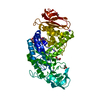 6tp1C 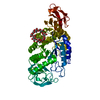 6tp2C S: Starting model for refinement C: citing same article ( |
|---|---|
| Similar structure data |


-Links
 PDBj
PDBj


- Assembly
| Deposited unit | 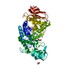
| ||||||||
|---|---|---|---|---|---|---|---|---|---|
| 1 |
| ||||||||
| Unit cell |
|
-Components
-Protein / Sugars , 2 types, 2 molecules A
| #1: Protein | Mass: 55313.863 Da / Num. of mol.: 1 Source method: isolated from a genetically manipulated source Source: (gene. exp.) |
|---|---|
| #2: Polysaccharide | alpha-D-glucopyranose-(1-4)-alpha-D-glucopyranose / alpha-maltose  Source method: isolated from a genetically manipulated source Details: oligosaccharide / References: alpha-maltose |
-Non-polymers , 5 types, 395 molecules 


| #3: Chemical |  Mass: 40.078 Da / Num. of mol.: 2 / Source method: obtained synthetically / Formula: Ca Mass: 40.078 Da / Num. of mol.: 2 / Source method: obtained synthetically / Formula: Ca#4: Chemical |  Mass: 22.990 Da / Num. of mol.: 2 / Source method: obtained synthetically / Formula: Na Mass: 22.990 Da / Num. of mol.: 2 / Source method: obtained synthetically / Formula: Na#5: Chemical | ChemComp-MLI / |  Mass: 102.046 Da / Num. of mol.: 1 / Source method: obtained synthetically / Formula: C3H2O4 Mass: 102.046 Da / Num. of mol.: 1 / Source method: obtained synthetically / Formula: C3H2O4#6: Chemical |  Mass: 60.052 Da / Num. of mol.: 2 / Source method: obtained synthetically / Formula: C2H4O2 Mass: 60.052 Da / Num. of mol.: 2 / Source method: obtained synthetically / Formula: C2H4O2#7: Water | ChemComp-HOH / | Mass: 18.015 Da / Num. of mol.: 388 / Source method: isolated from a natural source / Formula: H2O |
|---|
-Details
| Has ligand of interest | Y |
|---|
-Experimental details
-Experiment
| Experiment | Method: X-RAY DIFFRACTION / Number of used crystals: 1 |
|---|
- Sample preparation
| Crystal | Density Matthews: 3 Å3/Da / Density % sol: 59 % |
|---|---|
| Crystal grow | Temperature: 294 K / Method: vapor diffusion, hanging drop / pH: 6 / Details: 42-46 % Tacsimate, 10 mM CaCl2 |
-Data collection
| Diffraction | Mean temperature: 110 K / Serial crystal experiment: N |
|---|---|
| Diffraction source | Source: ROTATING ANODE / Type: BRUKER AXS MICROSTAR / Wavelength: 1.5418 Å |
| Detector | Type: MARRESEARCH / Detector: IMAGE PLATE / Date: Jan 16, 2017 |
| Radiation | Protocol: SINGLE WAVELENGTH / Monochromatic (M) / Laue (L): M / Scattering type: x-ray |
| Radiation wavelength | Wavelength: 1.5418 Å / Relative weight: 1 |
| Reflection | Resolution: 2.03→49.89 Å / Num. obs: 42384 / % possible obs: 98.2 % / Redundancy: 8.1 % / Biso Wilson estimate: 12.5 Å2 / CC1/2: 0.995 / Rmerge(I) obs: 0.157 / Rpim(I) all: 0.058 / Rrim(I) all: 0.168 / Net I/σ(I): 11.1 |
| Reflection shell | Resolution: 2.03→2.08 Å / Redundancy: 7.5 % / Rmerge(I) obs: 0.671 / Num. unique obs: 2385 / CC1/2: 0.805 / Rpim(I) all: 0.257 / Rrim(I) all: 0.72 / % possible all: 76.3 |
- Processing
| Software |
| ||||||||||||||||||||||||||||||||||||||||||||||||||||||||||||
|---|---|---|---|---|---|---|---|---|---|---|---|---|---|---|---|---|---|---|---|---|---|---|---|---|---|---|---|---|---|---|---|---|---|---|---|---|---|---|---|---|---|---|---|---|---|---|---|---|---|---|---|---|---|---|---|---|---|---|---|---|---|
| Refinement | Method to determine structure: FOURIER SYNTHESIS Starting model: 6TOY Resolution: 2.04→48.89 Å / Cor.coef. Fo:Fc: 0.961 / Cor.coef. Fo:Fc free: 0.946 / SU B: 6.442 / SU ML: 0.09 / Cross valid method: THROUGHOUT / σ(F): 0 / ESU R: 0.142 / ESU R Free: 0.126 Details: U VALUES : WITH TLS ADDED HYDROGENS HAVE BEEN ADDED IN THE RIDING POSITIONS
| ||||||||||||||||||||||||||||||||||||||||||||||||||||||||||||
| Solvent computation | Ion probe radii: 0.7 Å / Shrinkage radii: 0.7 Å / VDW probe radii: 1 Å | ||||||||||||||||||||||||||||||||||||||||||||||||||||||||||||
| Displacement parameters | Biso max: 76.69 Å2 / Biso mean: 21.808 Å2 / Biso min: 10.58 Å2
| ||||||||||||||||||||||||||||||||||||||||||||||||||||||||||||
| Refinement step | Cycle: final / Resolution: 2.04→48.89 Å
| ||||||||||||||||||||||||||||||||||||||||||||||||||||||||||||
| Refine LS restraints |
| ||||||||||||||||||||||||||||||||||||||||||||||||||||||||||||
| LS refinement shell | Resolution: 2.041→2.094 Å / Rfactor Rfree error: 0 / Total num. of bins used: 20
| ||||||||||||||||||||||||||||||||||||||||||||||||||||||||||||
| Refinement TLS params. | Method: refined / Origin x: -8.526 Å / Origin y: -32.216 Å / Origin z: 16.778 Å
|
