 Movie
Movie Controller

[English] 日本語
 Yorodumi
- PDB-1wpc: Crystal structure of maltohexaose-producing amylase complexed wit... -
+ Open data

- Basic information
| Entry | Database: PDB / ID: 1wpc | |||||||||
|---|---|---|---|---|---|---|---|---|---|---|
| Title | Crystal structure of maltohexaose-producing amylase complexed with pseudo-maltononaose | |||||||||
 Components Components | Glucan 1,4-alpha-maltohexaosidase | |||||||||
 Keywords Keywords | HYDROLASE / Maltohexaose-producing amylase / alpha-amylase / acarbose | |||||||||
| Function / homology |  Function and homology information Function and homology informationglucan 1,4-alpha-maltohexaosidase / glucan 1,4-alpha-maltohexaosidase activity / starch catabolic process / alpha-amylase activity / calcium ion binding / extracellular region Similarity search - Function | |||||||||
| Biological species |  | |||||||||
| Method |  X-RAY DIFFRACTION / SYNCHROTRON / MOLECULAR REPLACEMENT / Resolution: 1.9 Å X-RAY DIFFRACTION / SYNCHROTRON / MOLECULAR REPLACEMENT / Resolution: 1.9 Å | |||||||||
 Authors Authors | Kanai, R. / Haga, K. / Akiba, T. / Yamane, K. / Harata, K. | |||||||||
 Citation Citation | Journal: Biochemistry / Year: 2004 Title: Biochemical and crystallographic analyses of maltohexaose-producing amylase from alkalophilic Bacillus sp. 707 Authors: Kanai, R. / Haga, K. / Akiba, T. / Yamane, K. / Harata, K. #1: Journal: Appl.Microbiol.Biotechnol. / Year: 1988Title: Cloning of a gene for maltohexaose producing amylase of an alkalophilic Bacillus and hyper-production of the enzyme in Bacillus subtilis cells Authors: Kimura, K. / Tsukamoto, A. / Ishii, Y. / Takano, T. / Yamane, K. | |||||||||
| History |
|
- Structure visualization
| Structure viewer | Molecule: MolmilJmol/JSmol |
|---|
- Downloads & links
-Download
| PDBx/mmCIF format | 1wpc.cif.gz | 123.5 KB | Display | PDBx/mmCIF format |
|---|---|---|---|---|
| PDB format | pdb1wpc.ent.gz | 93.2 KB | Display | PDB format |
| PDBx/mmJSON format | 1wpc.json.gz | Tree view | PDBx/mmJSON format | |
| Others |  Other downloads Other downloads |
-Validation report
| Arichive directory | https://data.pdbj.org/pub/pdb/validation_reports/wp/1wpcftp://data.pdbj.org/pub/pdb/validation_reports/wp/1wpc | HTTPS FTP |
|---|
-Related structure data
| Related structure data |  1wp6SC S: Starting model for refinement C: citing same article ( |
|---|---|
| Similar structure data |


-Links
 PDBj
PDBj


- Assembly
| Deposited unit | 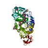
| ||||||||
|---|---|---|---|---|---|---|---|---|---|
| 1 |
| ||||||||
| Unit cell |
|
-Components
-Protein , 1 types, 1 molecules A
| #1: Protein | Mass: 55438.891 Da / Num. of mol.: 1 Source method: isolated from a genetically manipulated source Source: (gene. exp.) References: UniProt: P19571, glucan 1,4-alpha-maltohexaosidase |
|---|
-Sugars , 3 types, 4 molecules
| #2: Polysaccharide | 4,6-dideoxy-alpha-D-xylo-hexopyranose-(1-4)-alpha-D-glucopyranose-(1-4)-alpha-D-glucopyranose-(1-4)- ...4,6-dideoxy-alpha-D-xylo-hexopyranose-(1-4)-alpha-D-glucopyranose-(1-4)-alpha-D-glucopyranose-(1-4)-alpha-D-glucopyranose Source method: isolated from a genetically manipulated source |
|---|---|
| #3: Polysaccharide | 4,6-dideoxy-alpha-D-xylo-hexopyranose-(1-4)-alpha-D-glucopyranose-(1-4)-beta-D-galactopyranose Source method: isolated from a genetically manipulated source |
| #4: Polysaccharide |   Source method: isolated from a genetically manipulated source Details: oligosaccharide / References: alpha-maltose |
-Non-polymers , 4 types, 330 molecules 


| #5: Chemical |  Mass: 175.182 Da / Num. of mol.: 2 Mass: 175.182 Da / Num. of mol.: 2Source method: isolated from a genetically manipulated source Formula: C7H13NO4 #6: Chemical |  Mass: 40.078 Da / Num. of mol.: 3 / Source method: obtained synthetically / Formula: Ca Mass: 40.078 Da / Num. of mol.: 3 / Source method: obtained synthetically / Formula: Ca#7: Chemical | ChemComp-NA / |  Mass: 22.990 Da / Num. of mol.: 1 / Source method: obtained synthetically / Formula: Na Mass: 22.990 Da / Num. of mol.: 1 / Source method: obtained synthetically / Formula: Na#8: Water | ChemComp-HOH / | Mass: 18.015 Da / Num. of mol.: 324 / Source method: isolated from a natural source / Formula: H2O |
|---|
-Experimental details
-Experiment
| Experiment | Method: X-RAY DIFFRACTION / Number of used crystals: 1 |
|---|
- Sample preparation
| Crystal | Density Matthews: 2.1 Å3/Da / Density % sol: 41.4 % |
|---|---|
| Crystal grow | Temperature: 298 K / Method: vapor diffusion, hanging drop / pH: 8.5 Details: MPD, TrisHCl, Ammonium Phosphate, Calcium Chloride, Sodium Chloride, Acarbose, Maltotriose, pH 8.5, VAPOR DIFFUSION, HANGING DROP, temperature 298K |
-Data collection
| Diffraction | Mean temperature: 95 K |
|---|---|
| Diffraction source | Source: SYNCHROTRON / Site: Photon Factory  / Beamline: AR-NW12A / Wavelength: 1 Å / Beamline: AR-NW12A / Wavelength: 1 Å |
| Detector | Type: ADSC QUANTUM 210 / Detector: CCD / Date: Jun 3, 2003 |
| Radiation | Monochromator: Si 111 CHANNEL / Protocol: SINGLE WAVELENGTH / Monochromatic (M) / Laue (L): M / Scattering type: x-ray |
| Radiation wavelength | Wavelength: 1 Å / Relative weight: 1 |
| Reflection | Resolution: 1.9→50 Å / Num. all: 39906 / Num. obs: 39475 / % possible obs: 98.8 % / Observed criterion σ(I): 27.9 / Biso Wilson estimate: 13.8 Å2 / Rmerge(I) obs: 0.053 |
| Reflection shell | Resolution: 1.9→1.94 Å / Rmerge(I) obs: 0.106 / % possible all: 98.8 |
- Processing
| Software |
| ||||||||||||||||||||||||||||||||||||||||||||||||||||||||||||
|---|---|---|---|---|---|---|---|---|---|---|---|---|---|---|---|---|---|---|---|---|---|---|---|---|---|---|---|---|---|---|---|---|---|---|---|---|---|---|---|---|---|---|---|---|---|---|---|---|---|---|---|---|---|---|---|---|---|---|---|---|---|
| Refinement | Method to determine structure: MOLECULAR REPLACEMENT Starting model: PDB ENTRY 1WP6 Resolution: 1.9→6 Å / Rfactor Rfree error: 0.003 / Data cutoff high absF: 834402.78 / Data cutoff low absF: 0 / Isotropic thermal model: RESTRAINED / Cross valid method: THROUGHOUT / σ(F): 2 / Stereochemistry target values: Engh & Huber
| ||||||||||||||||||||||||||||||||||||||||||||||||||||||||||||
| Solvent computation | Solvent model: FLAT MODEL / Bsol: 93.1978 Å2 / ksol: 0.713919 e/Å3 | ||||||||||||||||||||||||||||||||||||||||||||||||||||||||||||
| Displacement parameters | Biso mean: 23.7 Å2
| ||||||||||||||||||||||||||||||||||||||||||||||||||||||||||||
| Refine analyze |
| ||||||||||||||||||||||||||||||||||||||||||||||||||||||||||||
| Refinement step | Cycle: LAST / Resolution: 1.9→6 Å
| ||||||||||||||||||||||||||||||||||||||||||||||||||||||||||||
| Refine LS restraints |
| ||||||||||||||||||||||||||||||||||||||||||||||||||||||||||||
| LS refinement shell | Refine-ID: X-RAY DIFFRACTION / Total num. of bins used: 6
| ||||||||||||||||||||||||||||||||||||||||||||||||||||||||||||
| Xplor file |
|
