| Entry | Database: PDB / ID: 6il3
|
|---|
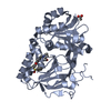
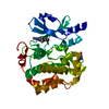
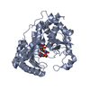
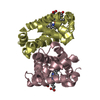
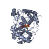
| Title | Crystal structure of the FLT3 kinase bound to a small molecule inhibitor |
|---|
 Components Components | Receptor-type tyrosine-protein kinase FLT3 |
|---|
 Keywords Keywords | TRANSFERASE / receptor tyrosine kinase type I inhibitor ATP binding domain DFG-in / SIGNALING PROTEIN |
|---|
| Function / homology |  Function and homology information Function and homology information
FLT3 mutants bind TKIs / KW2449-resistant FLT3 mutants / semaxanib-resistant FLT3 mutants / crenolanib-resistant FLT3 mutants / gilteritinib-resistant FLT3 mutants / lestaurtinib-resistant FLT3 mutants / midostaurin-resistant FLT3 mutants / pexidartinib-resistant FLT3 mutants / ponatinib-resistant FLT3 mutants / quizartinib-resistant FLT3 mutants ...FLT3 mutants bind TKIs / KW2449-resistant FLT3 mutants / semaxanib-resistant FLT3 mutants / crenolanib-resistant FLT3 mutants / gilteritinib-resistant FLT3 mutants / lestaurtinib-resistant FLT3 mutants / midostaurin-resistant FLT3 mutants / pexidartinib-resistant FLT3 mutants / ponatinib-resistant FLT3 mutants / quizartinib-resistant FLT3 mutants / sorafenib-resistant FLT3 mutants / sunitinib-resistant FLT3 mutants / tandutinib-resistant FLT3 mutants / linifanib-resistant FLT3 mutants / tamatinib-resistant FLT3 mutants / leukocyte homeostasis / lymphocyte proliferation / common myeloid progenitor cell proliferation / pro-B cell differentiation / dendritic cell differentiation / vascular endothelial growth factor receptor activity / nuclear glucocorticoid receptor binding / STAT5 Activation / phosphatidylinositol 3-kinase activator activity / myeloid progenitor cell differentiation / FLT3 signaling through SRC family kinases / cytokine receptor activity / cellular response to glucocorticoid stimulus / positive regulation of tyrosine phosphorylation of STAT protein / STAT5 activation downstream of FLT3 ITD mutants / growth factor binding / cellular response to cytokine stimulus / positive regulation of MAP kinase activity / hemopoiesis / PI3K Cascade / Signaling by FLT3 ITD and TKD mutants / peptidyl-tyrosine phosphorylation / transmembrane receptor protein tyrosine kinase activity / FLT3 Signaling / B cell differentiation / liver regeneration / FLT3 signaling by CBL mutants / Negative regulation of FLT3 / cell surface receptor protein tyrosine kinase signaling pathway / receptor protein-tyrosine kinase / Constitutive Signaling by Aberrant PI3K in Cancer / cytokine-mediated signaling pathway / PIP3 activates AKT signaling / protein autophosphorylation / cell migration / PI5P, PP2A and IER3 Regulate PI3K/AKT Signaling / RAF/MAP kinase cascade / protein tyrosine kinase activity / regulation of apoptotic process / positive regulation of MAPK cascade / positive regulation of phosphatidylinositol 3-kinase/protein kinase B signal transduction / signaling receptor complex / endosome membrane / endoplasmic reticulum lumen / positive regulation of cell population proliferation / protein-containing complex binding / endoplasmic reticulum / ATP binding / plasma membraneSimilarity search - Function Tyrosine-protein kinase, receptor class III, conserved site / Receptor tyrosine kinase class III signature. / Immunoglobulin / Immunoglobulin domain / : / Tyrosine-protein kinase, catalytic domain / Tyrosine kinase, catalytic domain / Tyrosine protein kinases specific active-site signature. / Tyrosine-protein kinase, active site / Protein tyrosine and serine/threonine kinase ...Tyrosine-protein kinase, receptor class III, conserved site / Receptor tyrosine kinase class III signature. / Immunoglobulin / Immunoglobulin domain / : / Tyrosine-protein kinase, catalytic domain / Tyrosine kinase, catalytic domain / Tyrosine protein kinases specific active-site signature. / Tyrosine-protein kinase, active site / Protein tyrosine and serine/threonine kinase / Serine-threonine/tyrosine-protein kinase, catalytic domain / Phosphorylase Kinase; domain 1 / Phosphorylase Kinase; domain 1 / Transferase(Phosphotransferase) domain 1 / Transferase(Phosphotransferase); domain 1 / Ig-like domain profile. / Immunoglobulin-like domain / Immunoglobulin-like domain superfamily / Protein kinase, ATP binding site / Protein kinases ATP-binding region signature. / Immunoglobulin-like fold / Protein kinase domain profile. / Protein kinase domain / Protein kinase-like domain superfamily / 2-Layer Sandwich / Orthogonal Bundle / Mainly Alpha / Alpha BetaSimilarity search - Domain/homology |
|---|
| Biological species |  Homo sapiens (human) Homo sapiens (human) |
|---|
| Method |  X-RAY DIFFRACTION / SYNCHROTRON / Resolution: 2.5 Å X-RAY DIFFRACTION / SYNCHROTRON / Resolution: 2.5 Å |
|---|
 Authors Authors | Thomas, C.J. |
|---|
 Citation Citation | Journal: To Be Published
Title: Crystal structure of the FLT3 kinase bound to a small molecule inhibitor
Authors: Thomas, C.J. |
|---|
| History | | Deposition | Oct 16, 2018 | Deposition site: PDBJ / Processing site: PDBJ |
|---|
| Revision 1.0 | Dec 12, 2018 | Provider: repository / Type: Initial release |
|---|
| Revision 1.1 | Mar 27, 2024 | Group: Data collection / Database references / Category: chem_comp_atom / chem_comp_bond / database_2
Item: _database_2.pdbx_DOI / _database_2.pdbx_database_accession |
|---|
|
|---|
 Movie
Movie Controller

 Yorodumi
 Open data
 Basic information
 Structure visualization
 Downloads & links
 Other downloads
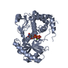


 PDBj
PDBj


 Assembly
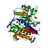
 Baculovirus expression vector pFastBac1-HM


 Mass: 305.334 Da / Num. of mol.: 1 / Source method: obtained synthetically / Formula: C17H15N5O
 Mass: 94.971 Da / Num. of mol.: 4 / Source method: obtained synthetically / Formula: PO4
 Mass: 221.317 Da / Num. of mol.: 2 / Source method: obtained synthetically / Formula: C9H19NO3S / Comment: pH buffer*YM
 Mass: 92.094 Da / Num. of mol.: 10 / Source method: obtained synthetically / Formula: C3H8O3
 Mass: 35.453 Da / Num. of mol.: 3 / Source method: obtained synthetically / Formula: Cl
 Sample preparation
 / Beamline: BL18U1 / Wavelength: 0.979 Å
 Processing