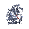
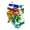
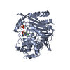
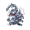
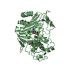
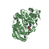
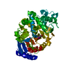
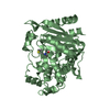
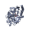
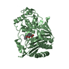
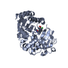
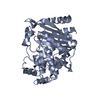
Journal: Acta Crystallogr D Struct Biol / Year: 2016 Title: Structural basis for the extended substrate spectrum of AmpC BER and structure-guided discovery of the inhibition activity of citrate against the class C beta-lactamases AmpC BER and CMY-10. Authors: Na, J.H. / Cha, S.S.
Resolution: 1.75→50 Å / Cor.coef. Fo:Fc: 0.963 / Cor.coef. Fo:Fc free: 0.954 / SU B: 2.156 / SU ML: 0.066 / Cross valid method: THROUGHOUT / ESU R: 0.085 / ESU R Free: 0.085 / Details: HYDROGENS HAVE BEEN ADDED IN THE RIDING POSITIONS
Rfactor
Num. reflection
% reflection
Selection details
Rfree
0.20015
5514
5 %
RANDOM
Rwork
0.17679
-
-
-
obs
0.17799
104247
96.51 %
-
Solvent computation
Ion probe radii: 0.8 Å / Shrinkage radii: 0.8 Å / VDW probe radii: 1.2 Å
 Movie
Movie Controller


 Open data
 Basic information
 Components
 Keywords
 Function and homology information
 X-RAY DIFFRACTION /
  Authors
 Korea, Republic Of, 1items
  Citation
 Structure visualization
 Downloads & links
 Other downloads


 PDBj
PDBj


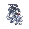
 Assembly


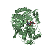
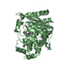
 Mass: 192.124 Da / Num. of mol.: 4 / Source method: obtained synthetically / Formula: C6H8O7
 Mass: 18.015 Da / Num. of mol.: 269 / Source method: isolated from a natural source / Formula: H2O
 Sample preparation
 Processing