 Movie
Movie Controller


+ Open data

- Basic information
| Entry | Database: PDB / ID: 6ijz | |||||||||
|---|---|---|---|---|---|---|---|---|---|---|
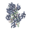
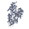
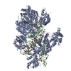
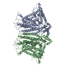
| Title | Structure of a plant cation channel | |||||||||
 Components Components | Calcium permeable stress-gated cation channel 1 | |||||||||
 Keywords Keywords | MEMBRANE PROTEIN / Channel | |||||||||
| Function / homology |  Function and homology information Function and homology informationmechanosensitive monoatomic ion channel activity / calcium-activated cation channel activity / monoatomic cation transport / identical protein binding / plasma membrane Similarity search - Function | |||||||||
| Biological species |  | |||||||||
| Method | ELECTRON MICROSCOPY / single particle reconstruction / cryo EM / Resolution: 3.68 Å | |||||||||
 Authors Authors | Sun, L. / Wang, J. / Liu, X. | |||||||||
| Funding support |  China, 2items China, 2items
| |||||||||
 Citation Citation | Journal: Nat Commun / Year: 2018 Title: Structure of the hyperosmolality-gated calcium-permeable channel OSCA1.2. Authors: Xin Liu / Jiawei Wang / Linfeng Sun / Abstract: In plants, hyperosmolality stimuli triggers opening of the osmosensitive channels, leading to a rapid downstream signaling cascade initiated by cytosolic calcium concentration elevation. Members of ...In plants, hyperosmolality stimuli triggers opening of the osmosensitive channels, leading to a rapid downstream signaling cascade initiated by cytosolic calcium concentration elevation. Members of the OSCA family in Arabidopsis thaliana, identified as the hyperosmolality-gated calcium-permeable channels, have been suggested to play a key role during the initial phase of hyperosmotic stress response. Here, we report the atomic structure of Arabidopsis OSCA1.2 determined by single-particle cryo-electron microscopy. It contains 11 transmembrane helices and forms a homodimer. It is in an inactivated state, and the pore-lining residues are clearly identified. Its cytosolic domain contains a RNA recognition motif and two unique long helices. The linker between these two helices forms an anchor in the lipid bilayer and may be essential to osmosensing. The structure of AtOSCA1.2 serves as a platform for the study of the mechanism underlying osmotic stress responses and mechanosensing. | |||||||||
| History |
|
- Structure visualization
| Movie |
Movie viewer |
|---|---|
| Structure viewer | Molecule: MolmilJmol/JSmol |
- Downloads & links
-Download
| PDBx/mmCIF format | 6ijz.cif.gz | 246.7 KB | Display | PDBx/mmCIF format |
|---|---|---|---|---|
| PDB format | pdb6ijz.ent.gz | 199.5 KB | Display | PDB format |
| PDBx/mmJSON format | 6ijz.json.gz | Tree view | PDBx/mmJSON format | |
| Others |  Other downloads Other downloads |
-Validation report
| Arichive directory | https://data.pdbj.org/pub/pdb/validation_reports/ij/6ijzftp://data.pdbj.org/pub/pdb/validation_reports/ij/6ijz | HTTPS FTP |
|---|
-Related structure data
| Related structure data |  9682MC  9677C M: map data used to model this data C: citing same article ( |
|---|---|
| Similar structure data |


-Links
 PDBj
PDBj- Assembly
| Deposited unit | 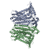
|
|---|---|
| 1 |
|
-Components
| #1: Protein | Mass: 87985.547 Da / Num. of mol.: 2 Source method: isolated from a genetically manipulated source Source: (gene. exp.)   Spodoptera frugiperda (fall armyworm) / References: UniProt: Q5XEZ5 Spodoptera frugiperda (fall armyworm) / References: UniProt: Q5XEZ5 |
|---|
-Experimental details
-Experiment
| Experiment | Method: ELECTRON MICROSCOPY |
|---|---|
| EM experiment | Aggregation state: PARTICLE / 3D reconstruction method: single particle reconstruction |
- Sample preparation
| Component | Name: Plant chennel / Type: CELL Details: Protein expressed in Sf-9 cell and purified in digitonin Entity ID: all / Source: MULTIPLE SOURCES |
|---|---|
| Source (natural) | Organism: |
| Source (recombinant) | Organism: Spodoptera frugiperda (fall armyworm) |
| Buffer solution | pH: 7.4 |
| Specimen | Conc.: 5 mg/ml / Embedding applied: NO / Shadowing applied: NO / Staining applied: NO / Vitrification applied: YES / Details: This sample was monodisperse. |
| Vitrification | Instrument: FEI VITROBOT MARK IV / Cryogen name: ETHANE / Humidity: 100 % / Chamber temperature: 281 K / Details: blot for 4 seconds before plunging |
- Electron microscopy imaging
| Experimental equipment |  Model: Titan Krios / Image courtesy: FEI Company |
|---|---|
| Microscopy | Model: FEI TITAN KRIOS |
| Electron gun | Electron source:  FIELD EMISSION GUN / Accelerating voltage: 300 kV / Illumination mode: SPOT SCAN FIELD EMISSION GUN / Accelerating voltage: 300 kV / Illumination mode: SPOT SCAN |
| Electron lens | Mode: BRIGHT FIELD |
| Specimen holder | Cryogen: NITROGEN / Specimen holder model: FEI TITAN KRIOS AUTOGRID HOLDER |
| Image recording | Average exposure time: 8 sec. / Electron dose: 50 e/Å2 / Detector mode: SUPER-RESOLUTION / Film or detector model: GATAN K2 SUMMIT (4k x 4k) |
| Image scans | Movie frames/image: 32 |
- Processing
| Software | Name: PHENIX / Version: 1.13_2998: / Classification: refinement | ||||||||||||||||||||||||||||||||||||
|---|---|---|---|---|---|---|---|---|---|---|---|---|---|---|---|---|---|---|---|---|---|---|---|---|---|---|---|---|---|---|---|---|---|---|---|---|---|
| EM software |
| ||||||||||||||||||||||||||||||||||||
| CTF correction | Type: PHASE FLIPPING AND AMPLITUDE CORRECTION | ||||||||||||||||||||||||||||||||||||
| Symmetry | Point symmetry: C2 (2 fold cyclic) | ||||||||||||||||||||||||||||||||||||
| 3D reconstruction | Resolution: 3.68 Å / Resolution method: FSC 0.143 CUT-OFF / Num. of particles: 271802 / Symmetry type: POINT | ||||||||||||||||||||||||||||||||||||
| Atomic model building | Protocol: AB INITIO MODEL / Space: REAL | ||||||||||||||||||||||||||||||||||||
| Refine LS restraints |
|
