 Movie
Movie Controller

[English] 日本語
 Yorodumi
- PDB-6ije: Crystal structure of the type VI amidase immunity (Tai4) from Agr... -
+ Open data

- Basic information
| Entry | Database: PDB / ID: 6ije | ||||||
|---|---|---|---|---|---|---|---|
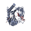
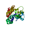
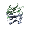
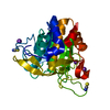
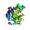
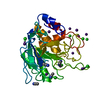
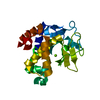
| Title | Crystal structure of the type VI amidase immunity (Tai4) from Agrobacterium tumefaciens | ||||||
 Components Components | Tai4 | ||||||
 Keywords Keywords | ANTITOXIN | ||||||
| Function / homology | Rap1a immunity protein / Rap1a immunity proteins / Rap1a domain-containing protein Function and homology information Function and homology information | ||||||
| Biological species |  Rhizobium radiobacter (Agrobacterium genomosp. 4) Rhizobium radiobacter (Agrobacterium genomosp. 4) | ||||||
| Method |  X-RAY DIFFRACTION / SYNCHROTRON / MOLECULAR REPLACEMENT / Resolution: 1.55 Å X-RAY DIFFRACTION / SYNCHROTRON / MOLECULAR REPLACEMENT / Resolution: 1.55 Å | ||||||
 Authors Authors | Fukuhara, S. / Nakane, T. / Yamashita, K. / Ishii, R. / Ishitani, R. / Nureki, O. | ||||||
 Citation Citation | Journal: Acta Crystallogr F Struct Biol Commun / Year: 2018 Title: Crystal structure of the Agrobacterium tumefaciens type VI effector-immunity complex. Authors: Fukuhara, S. / Nakane, T. / Yamashita, K. / Ishii, R. / Ishitani, R. / Nureki, O. | ||||||
| History |
|
- Structure visualization
| Structure viewer | Molecule: MolmilJmol/JSmol |
|---|
- Downloads & links
-Download
| PDBx/mmCIF format | 6ije.cif.gz | 56.7 KB | Display | PDBx/mmCIF format |
|---|---|---|---|---|
| PDB format | pdb6ije.ent.gz | 39.9 KB | Display | PDB format |
| PDBx/mmJSON format | 6ije.json.gz | Tree view | PDBx/mmJSON format | |
| Others |  Other downloads Other downloads |
-Validation report
| Arichive directory | https://data.pdbj.org/pub/pdb/validation_reports/ij/6ijeftp://data.pdbj.org/pub/pdb/validation_reports/ij/6ije | HTTPS FTP |
|---|
-Related structure data
| Related structure data | 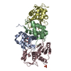 6ijfC 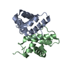 3zfiS S: Starting model for refinement C: citing same article ( |
|---|---|
| Similar structure data |


-Links
 PDBj
PDBj- Assembly
| Deposited unit | 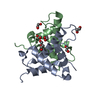
| ||||||||||||||||||
|---|---|---|---|---|---|---|---|---|---|---|---|---|---|---|---|---|---|---|---|
| 1 |
| ||||||||||||||||||
| Unit cell |
| ||||||||||||||||||
| Noncrystallographic symmetry (NCS) | NCS domain:
NCS domain segments: Component-ID: _ / Ens-ID: 1 / Beg auth comp-ID: GLU / Beg label comp-ID: GLU / End auth comp-ID: CYS / End label comp-ID: CYS / Refine code: _ / Auth seq-ID: 2 - 99 / Label seq-ID: 7 - 104
|
-Components
| #1: Protein | Mass: 11694.150 Da / Num. of mol.: 2 Source method: isolated from a genetically manipulated source Source: (gene. exp.) Rhizobium radiobacter (Agrobacterium genomosp. 4)Gene: SY94_4314 / Plasmid: pCold-GST / Production host: #2: Chemical | ChemComp-EDO /   Mass: 62.068 Da / Num. of mol.: 7 / Source method: obtained synthetically / Formula: C2H6O2 Mass: 62.068 Da / Num. of mol.: 7 / Source method: obtained synthetically / Formula: C2H6O2#3: Water | ChemComp-HOH / |  Mass: 18.015 Da / Num. of mol.: 141 / Source method: isolated from a natural source / Formula: H2O Mass: 18.015 Da / Num. of mol.: 141 / Source method: isolated from a natural source / Formula: H2OHas protein modification | Y | |
|---|
-Experimental details
-Experiment
| Experiment | Method: X-RAY DIFFRACTION / Number of used crystals: 1 |
|---|
- Sample preparation
| Crystal | Density Matthews: 2.38 Å3/Da / Density % sol: 48.3 % / Mosaicity: 0 ° |
|---|---|
| Crystal grow | Temperature: 293 K / Method: vapor diffusion, sitting drop / pH: 8 Details: 33 % PEG 6000, 1.5 M lithium chloride, 100 mM sodium acetate |
-Data collection
| Diffraction | Mean temperature: 100 K / Serial crystal experiment: N | ||||||||||||||||||||||||
|---|---|---|---|---|---|---|---|---|---|---|---|---|---|---|---|---|---|---|---|---|---|---|---|---|---|
| Diffraction source | Source: SYNCHROTRON / Site: SPring-8  / Beamline: BL41XU / Wavelength: 1 Å / Beamline: BL41XU / Wavelength: 1 Å | ||||||||||||||||||||||||
| Detector | Type: DECTRIS PILATUS3 6M / Detector: PIXEL / Date: May 19, 2015 | ||||||||||||||||||||||||
| Radiation | Monochromator: Si(111) / Protocol: SINGLE WAVELENGTH / Monochromatic (M) / Laue (L): M / Scattering type: x-ray | ||||||||||||||||||||||||
| Radiation wavelength | Wavelength: 1 Å / Relative weight: 1 | ||||||||||||||||||||||||
| Reflection | Resolution: 1.55→53.92 Å / Num. obs: 32919 / % possible obs: 99.5 % / Redundancy: 6.2 % / CC1/2: 0.998 / Rmerge(I) obs: 0.067 / Rpim(I) all: 0.029 / Rrim(I) all: 0.073 / Net I/σ(I): 13 / Num. measured all: 203509 / Scaling rejects: 9 | ||||||||||||||||||||||||
| Reflection shell | Diffraction-ID: 1
|
- Processing
| Software |
| ||||||||||||||||||||||||||||||||||||||||||||||||||||||||||||
|---|---|---|---|---|---|---|---|---|---|---|---|---|---|---|---|---|---|---|---|---|---|---|---|---|---|---|---|---|---|---|---|---|---|---|---|---|---|---|---|---|---|---|---|---|---|---|---|---|---|---|---|---|---|---|---|---|---|---|---|---|---|
| Refinement | Method to determine structure: MOLECULAR REPLACEMENT Starting model: 3ZFI Resolution: 1.55→44.96 Å / Cor.coef. Fo:Fc: 0.968 / Cor.coef. Fo:Fc free: 0.963 / SU B: 1.375 / SU ML: 0.049 / Cross valid method: FREE R-VALUE / σ(F): 0 / ESU R: 0.073 / ESU R Free: 0.073 Details: HYDROGENS HAVE BEEN ADDED IN THE RIDING POSITIONS U VALUES : REFINED INDIVIDUALLY
| ||||||||||||||||||||||||||||||||||||||||||||||||||||||||||||
| Solvent computation | Ion probe radii: 0.8 Å / Shrinkage radii: 0.8 Å / VDW probe radii: 1.2 Å | ||||||||||||||||||||||||||||||||||||||||||||||||||||||||||||
| Displacement parameters | Biso max: 98.96 Å2 / Biso mean: 25.599 Å2 / Biso min: 13.78 Å2
| ||||||||||||||||||||||||||||||||||||||||||||||||||||||||||||
| Refinement step | Cycle: final / Resolution: 1.55→44.96 Å
| ||||||||||||||||||||||||||||||||||||||||||||||||||||||||||||
| Refine LS restraints |
| ||||||||||||||||||||||||||||||||||||||||||||||||||||||||||||
| Refine LS restraints NCS | Ens-ID: 1 / Number: 3044 / Refine-ID: X-RAY DIFFRACTION / Type: interatomic distance / Rms dev position: 0.14 Å / Weight position: 0.01
| ||||||||||||||||||||||||||||||||||||||||||||||||||||||||||||
| LS refinement shell | Resolution: 1.55→1.59 Å / Rfactor Rfree error: 0 / Total num. of bins used: 20
|
