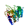
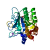
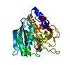
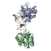
[English] 日本語
Yorodumi
- PDB-2wuv: Crystallographic analysis of counter-ion effects on subtilisin en... -

+
Open data


ID or keywords:

Loading...

-
Basic information

Entry
Database: PDB / ID: 2wuv
TitleCrystallographic analysis of counter-ion effects on subtilisin enzymatic action in acetonitrile
ComponentsSUBTILISIN CARLSBERG
KeywordsHYDROLASE / SERINE PROTEASE / METAL-BINDING
Function / homology
Function and homology information


subtilisin / serine-type endopeptidase activity / proteolysis / extracellular space / metal ion binding
Similarity search - Function
Subtilisin Carlsberg-like catalytic domain / Peptidase S8/S53 domain / Peptidase S8 propeptide/proteinase inhibitor I9 / Peptidase inhibitor I9 / Peptidase S8 propeptide/proteinase inhibitor I9 superfamily / Peptidase S8, subtilisin, His-active site / Serine proteases, subtilase family, histidine active site. / Serine proteases, subtilase family, aspartic acid active site. / Peptidase S8, subtilisin, Asp-active site / Serine proteases, subtilase family, serine active site. ...Subtilisin Carlsberg-like catalytic domain / Peptidase S8/S53 domain / Peptidase S8 propeptide/proteinase inhibitor I9 / Peptidase inhibitor I9 / Peptidase S8 propeptide/proteinase inhibitor I9 superfamily / Peptidase S8, subtilisin, His-active site / Serine proteases, subtilase family, histidine active site. / Serine proteases, subtilase family, aspartic acid active site. / Peptidase S8, subtilisin, Asp-active site / Serine proteases, subtilase family, serine active site. / Peptidase S8, subtilisin, Ser-active site / Serine proteases, subtilase domain profile. / Peptidase S8, subtilisin-related / Peptidase S8/S53 domain superfamily / Subtilase family / Peptidase S8/S53 domain / Rossmann fold / 3-Layer(aba) Sandwich / Alpha Beta
Similarity search - Domain/homology
ACETONITRILE / : / Subtilisin Carlsberg
Similarity search - Component
Biological speciesBACILLUS LICHENIFORMIS (bacteria)
MethodX-RAY DIFFRACTION / SYNCHROTRON / MOLECULAR REPLACEMENT / Resolution: 2.24 Å
AuthorsCianci, M. / Tomaszewki, B. / Helliwell, J.R. / Halling, P.J.
CitationJournal: J.Am.Chem.Soc. / Year: 2010
Title: Crystallographic Analysis of Counterion Effects on Subtilisin Enzymatic Action in Acetonitrile.
Authors: Cianci, M. / Tomaszewski, B. / Helliwell, J.R. / Halling, P.J.
History
DepositionOct 9, 2009Deposition site: PDBE / Processing site: PDBE
Revision 1.0Dec 8, 2010Provider: repository / Type: Initial release
Revision 1.1May 8, 2011Group: Version format compliance
Revision 1.2Jul 13, 2011Group: Version format compliance
Revision 1.3Dec 20, 2023Group: Data collection / Database references ...Data collection / Database references / Derived calculations / Other / Refinement description
Category: chem_comp_atom / chem_comp_bond ...chem_comp_atom / chem_comp_bond / database_2 / pdbx_database_status / pdbx_initial_refinement_model / pdbx_struct_conn_angle / struct_conn / struct_site
Item: _database_2.pdbx_DOI / _database_2.pdbx_database_accession ..._database_2.pdbx_DOI / _database_2.pdbx_database_accession / _pdbx_database_status.status_code_sf / _pdbx_struct_conn_angle.ptnr1_auth_comp_id / _pdbx_struct_conn_angle.ptnr1_auth_seq_id / _pdbx_struct_conn_angle.ptnr1_label_asym_id / _pdbx_struct_conn_angle.ptnr1_label_atom_id / _pdbx_struct_conn_angle.ptnr1_label_comp_id / _pdbx_struct_conn_angle.ptnr1_label_seq_id / _pdbx_struct_conn_angle.ptnr1_symmetry / _pdbx_struct_conn_angle.ptnr2_auth_comp_id / _pdbx_struct_conn_angle.ptnr2_auth_seq_id / _pdbx_struct_conn_angle.ptnr2_label_asym_id / _pdbx_struct_conn_angle.ptnr2_label_atom_id / _pdbx_struct_conn_angle.ptnr2_label_comp_id / _pdbx_struct_conn_angle.ptnr3_auth_comp_id / _pdbx_struct_conn_angle.ptnr3_auth_seq_id / _pdbx_struct_conn_angle.ptnr3_label_asym_id / _pdbx_struct_conn_angle.ptnr3_label_atom_id / _pdbx_struct_conn_angle.ptnr3_label_comp_id / _pdbx_struct_conn_angle.ptnr3_label_seq_id / _pdbx_struct_conn_angle.ptnr3_symmetry / _pdbx_struct_conn_angle.value / _struct_conn.pdbx_dist_value / _struct_conn.ptnr1_auth_comp_id / _struct_conn.ptnr1_auth_seq_id / _struct_conn.ptnr1_label_asym_id / _struct_conn.ptnr1_label_atom_id / _struct_conn.ptnr1_label_comp_id / _struct_conn.ptnr1_label_seq_id / _struct_conn.ptnr1_symmetry / _struct_conn.ptnr2_auth_comp_id / _struct_conn.ptnr2_auth_seq_id / _struct_conn.ptnr2_label_asym_id / _struct_conn.ptnr2_label_atom_id / _struct_conn.ptnr2_label_comp_id / _struct_conn.ptnr2_label_seq_id / _struct_conn.ptnr2_symmetry / _struct_site.pdbx_auth_asym_id / _struct_site.pdbx_auth_comp_id / _struct_site.pdbx_auth_seq_id

-
Structure visualization

Structure viewerMolecule:
MolmilJmol/JSmol

Downloads & links

-
Assembly

Deposited unit
A: SUBTILISIN CARLSBERG
hetero molecules


Theoretical massNumber of molelcules
Total (without water)29,32027
Polymers27,3061
Non-polymers2,01426
Water2,036113
1


  • Idetical with deposited unit
  • defined by author&software
TypeNameSymmetry operationNumber
identity operation1_555x,y,z1
MethodPISA
Unit cell
Length a, b, c (Å)52.177, 55.311, 75.778
Angle α, β, γ (deg.)90.00, 90.00, 90.00
Int Tables number19
Space group name H-MP212121

-
Components

-
Protein , 1 types, 1 molecules A

#1: Protein SUBTILISIN CARLSBERG


Mass: 27306.199 Da / Num. of mol.: 1 / Fragment: RESIDUES 106-379 / Source method: isolated from a natural source / Source: (natural) BACILLUS LICHENIFORMIS (bacteria) / References: UniProt: P00780, subtilisin

-
Non-polymers , 6 types, 139 molecules

#2: Chemical
ChemComp-CL / CHLORIDE ION


Mass: 35.453 Da / Num. of mol.: 8 / Source method: obtained synthetically / Formula: Cl
#3: Chemical
ChemComp-CS / CESIUM ION


Mass: 132.905 Da / Num. of mol.: 11 / Source method: obtained synthetically / Formula: Cs
#4: Chemical ChemComp-CA / CALCIUM ION


Mass: 40.078 Da / Num. of mol.: 1 / Source method: obtained synthetically / Formula: Ca
#5: Chemical ChemComp-NA / SODIUM ION


Mass: 22.990 Da / Num. of mol.: 1 / Source method: obtained synthetically / Formula: Na
#6: Chemical
ChemComp-CCN / ACETONITRILE


Mass: 41.052 Da / Num. of mol.: 5 / Source method: obtained synthetically / Formula: C2H3N
#7: Water ChemComp-HOH / water


Mass: 18.015 Da / Num. of mol.: 113 / Source method: isolated from a natural source / Formula: H2O

-
Experimental details

-
Experiment

ExperimentMethod: X-RAY DIFFRACTION

-
Sample preparation

CrystalDensity Matthews: 1.89 Å3/Da / Density % sol: 30.4 % / Description: NONE
Crystal growDetails: SUBTILISIN WAS CRYSTALLIZED BY THE BATCH METHOD FROM BUFFER SOLUTION SATURATED WITH NA2SO4 ABOUT 13% (W/V) AS PRECIPITANT (32 PETSKO AND TSERNOGLOU 1976)

-
Data collection

DiffractionMean temperature: 287 K
Diffraction sourceSource: SYNCHROTRON / Site: SRS / Beamline: PX10.1 / Wavelength: 2.167
DetectorType: MARRESEARCH / Detector: CCD
RadiationMonochromator: SI 111 / Protocol: SINGLE WAVELENGTH / Monochromatic (M) / Laue (L): M / Scattering type: x-ray
Radiation wavelengthWavelength: 2.167 Å / Relative weight: 1
ReflectionResolution: 2.24→44.68 Å / Num. obs: 10215 / % possible obs: 97.4 % / Observed criterion σ(I): 2 / Redundancy: 26.4 % / Rmerge(I) obs: 0.2 / Net I/σ(I): 14.1
Reflection shellResolution: 2.24→2.29 Å / Redundancy: 9.9 % / Rmerge(I) obs: 0.22 / Mean I/σ(I) obs: 8.4 / % possible all: 94.5

-
Processing

Software
NameVersionClassification
REFMAC5.5.0066refinement
HKL-2000data reduction
HKL-2000data scaling
MOLREPphasing
RefinementMethod to determine structure: MOLECULAR REPLACEMENT
Starting model: PDB ENTRY 1SCA
Resolution: 2.24→44.68 Å / Cor.coef. Fo:Fc: 0.958 / Cor.coef. Fo:Fc free: 0.923 / SU B: 5.286 / SU ML: 0.136 / Cross valid method: THROUGHOUT / ESU R: 0.38 / ESU R Free: 0.238 / Stereochemistry target values: MAXIMUM LIKELIHOOD / Details: HYDROGENS HAVE BEEN ADDED IN THE RIDING POSITIONS.
RfactorNum. reflection% reflectionSelection details
Rfree0.23054 515 4.8 %RANDOM
Rwork0.16 ---
obs0.1634 10215 97.37 %-
Solvent computationIon probe radii: 0.8 Å / Shrinkage radii: 0.8 Å / VDW probe radii: 1.4 Å / Solvent model: MASK
Displacement parametersBiso mean: 25.357 Å2
Baniso -1Baniso -2Baniso -3
1--0.14 Å20 Å20 Å2
2---1.82 Å20 Å2
3---1.95 Å2
Refinement stepCycle: LAST / Resolution: 2.24→44.68 Å
ProteinNucleic acidLigandSolventTotal
Num. atoms1920 0 36 113 2069
Refine LS restraints
Refine-IDTypeDev idealDev ideal targetNumber
X-RAY DIFFRACTIONr_bond_refined_d0.0190.0221961
X-RAY DIFFRACTIONr_bond_other_d
X-RAY DIFFRACTIONr_angle_refined_deg1.81.9392666
X-RAY DIFFRACTIONr_angle_other_deg
X-RAY DIFFRACTIONr_dihedral_angle_1_deg5.945272
X-RAY DIFFRACTIONr_dihedral_angle_2_deg34.0525.37367
X-RAY DIFFRACTIONr_dihedral_angle_3_deg15.88215272
X-RAY DIFFRACTIONr_dihedral_angle_4_deg15.753154
X-RAY DIFFRACTIONr_chiral_restr0.1240.2315
X-RAY DIFFRACTIONr_gen_planes_refined0.0090.0211480
X-RAY DIFFRACTIONr_gen_planes_other
X-RAY DIFFRACTIONr_nbd_refined
X-RAY DIFFRACTIONr_nbd_other
X-RAY DIFFRACTIONr_nbtor_refined
X-RAY DIFFRACTIONr_nbtor_other
X-RAY DIFFRACTIONr_xyhbond_nbd_refined
X-RAY DIFFRACTIONr_xyhbond_nbd_other
X-RAY DIFFRACTIONr_metal_ion_refined
X-RAY DIFFRACTIONr_metal_ion_other
X-RAY DIFFRACTIONr_symmetry_vdw_refined
X-RAY DIFFRACTIONr_symmetry_vdw_other
X-RAY DIFFRACTIONr_symmetry_hbond_refined
X-RAY DIFFRACTIONr_symmetry_hbond_other
X-RAY DIFFRACTIONr_symmetry_metal_ion_refined
X-RAY DIFFRACTIONr_symmetry_metal_ion_other
X-RAY DIFFRACTIONr_mcbond_it0.9741.51348
X-RAY DIFFRACTIONr_mcbond_other
X-RAY DIFFRACTIONr_mcangle_it1.7622134
X-RAY DIFFRACTIONr_mcangle_other
X-RAY DIFFRACTIONr_scbond_it3.1353613
X-RAY DIFFRACTIONr_scbond_other
X-RAY DIFFRACTIONr_scangle_it5.0724.5532
X-RAY DIFFRACTIONr_scangle_other
X-RAY DIFFRACTIONr_long_range_B_refined
X-RAY DIFFRACTIONr_long_range_B_other
X-RAY DIFFRACTIONr_rigid_bond_restr
X-RAY DIFFRACTIONr_sphericity_free
X-RAY DIFFRACTIONr_sphericity_bonded
LS refinement shellResolution: 2.24→2.298 Å / Total num. of bins used: 20
RfactorNum. reflection% reflection
Rfree0.34 34 -
Rwork0.278 707 -
obs--94.64 %

+
About Yorodumi

-
News

-
Feb 9, 2022. New format data for meta-information of EMDB entries

New format data for meta-information of EMDB entries

  • Version 3 of the EMDB header file is now the official format.
  • The previous official version 1.9 will be removed from the archive.

Related info.:EMDB header

External links:wwPDB to switch to version 3 of the EMDB data model

-
Aug 12, 2020. Covid-19 info

Covid-19 info

URL: https://pdbjlvh1.pdbj.org/emnavi/covid19.php

New page: Covid-19 featured information page in EM Navigator.

Related info.:Covid-19 info / Mar 5, 2020. Novel coronavirus structure data

+
Mar 5, 2020. Novel coronavirus structure data

Novel coronavirus structure data

Related info.:Yorodumi Speices / Aug 12, 2020. Covid-19 info

External links:COVID-19 featured content - PDBj / Molecule of the Month (242):Coronavirus Proteases

+
Jan 31, 2019. EMDB accession codes are about to change! (news from PDBe EMDB page)

EMDB accession codes are about to change! (news from PDBe EMDB page)

  • The allocation of 4 digits for EMDB accession codes will soon come to an end. Whilst these codes will remain in use, new EMDB accession codes will include an additional digit and will expand incrementally as the available range of codes is exhausted. The current 4-digit format prefixed with “EMD-” (i.e. EMD-XXXX) will advance to a 5-digit format (i.e. EMD-XXXXX), and so on. It is currently estimated that the 4-digit codes will be depleted around Spring 2019, at which point the 5-digit format will come into force.
  • The EM Navigator/Yorodumi systems omit the EMD- prefix.

Related info.:Q: What is EMD? / ID/Accession-code notation in Yorodumi/EM Navigator

External links:EMDB Accession Codes are Changing Soon! / Contact to PDBj

+
Jul 12, 2017. Major update of PDB

Major update of PDB

  • wwPDB released updated PDB data conforming to the new PDBx/mmCIF dictionary.
  • This is a major update changing the version number from 4 to 5, and with Remediation, in which all the entries are updated.
  • In this update, many items about electron microscopy experimental information are reorganized (e.g. em_software).
  • Now, EM Navigator and Yorodumi are based on the updated data.

External links:wwPDB Remediation / Enriched Model Files Conforming to OneDep Data Standards Now Available in the PDB FTP Archive

-
Yorodumi

Thousand views of thousand structures

  • Yorodumi is a browser for structure data from EMDB, PDB, SASBDB, etc.
  • This page is also the successor to EM Navigator detail page, and also detail information page/front-end page for Omokage search.
  • The word "yorodu" (or yorozu) is an old Japanese word meaning "ten thousand". "mi" (miru) is to see.

Related info.:EMDB / PDB / SASBDB / Comparison of 3 databanks / Yorodumi Search / Aug 31, 2016. New EM Navigator & Yorodumi / Yorodumi Papers / Jmol/JSmol / Function and homology information / Changes in new EM Navigator and Yorodumi

Read more