| Entry | Database: PDB / ID: 6ien
|
|---|
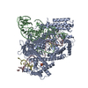
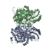
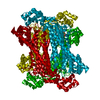
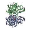
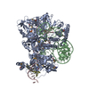
| Title | Substrate/product bound Argininosuccinate lyase from Mycobacterium tuberculosis |
|---|
 Components Components | Argininosuccinate lyase |
|---|
 Keywords Keywords | LYASE / arginine biosynthesis / tetramer / aspartase/fumarase / concerted movement |
|---|
| Function / homology |  Function and homology information Function and homology information
Argininosuccinate lyase / Argininosuccinate lyase, C-terminal / Argininosuccinate lyase C-terminal / Fumarase/aspartase (C-terminal domain) / Fumarate lyase, conserved site / Fumarate lyases signature. / Fumarase/aspartase (N-terminal domain) / Ribonucleotide Reductase Protein R1; domain 1 / Fumarate lyase family / Fumarate lyase, N-terminal ...Argininosuccinate lyase / Argininosuccinate lyase, C-terminal / Argininosuccinate lyase C-terminal / Fumarase/aspartase (C-terminal domain) / Fumarate lyase, conserved site / Fumarate lyases signature. / Fumarase/aspartase (N-terminal domain) / Ribonucleotide Reductase Protein R1; domain 1 / Fumarate lyase family / Fumarate lyase, N-terminal / Lyase / Fumarase/aspartase (Central domain) / Fumarase C; Chain A, domain 2 / Fumarase C; Chain B, domain 1 / Fumarase/histidase, N-terminal / L-Aspartase-like / Up-down Bundle / Orthogonal Bundle / Mainly AlphaSimilarity search - Domain/homology |
|---|
| Biological species |   Mycobacterium tuberculosis (bacteria) Mycobacterium tuberculosis (bacteria) |
|---|
| Method |  X-RAY DIFFRACTION / MOLECULAR REPLACEMENT / Resolution: 2.7 Å X-RAY DIFFRACTION / MOLECULAR REPLACEMENT / Resolution: 2.7 Å |
|---|
 Authors Authors | Paul, A. / Mishra, A. / Surolia, A. / Vijayan, M. |
|---|
 Citation Citation | Journal: IUBMB Life / Year: 2019
Title: Structural studies on M. tuberculosis argininosuccinate lyase and its liganded complex: Insights into catalytic mechanism.
Authors: Paul, A. / Mishra, A. / Surolia, A. / Vijayan, M. |
|---|
| History | | Deposition | Sep 14, 2018 | Deposition site: PDBJ / Processing site: PDBJ |
|---|
| Revision 1.0 | Feb 20, 2019 | Provider: repository / Type: Initial release |
|---|
| Revision 1.1 | Apr 17, 2019 | Group: Data collection / Database references / Category: citation / citation_author
Item: _citation.journal_volume / _citation.page_first ..._citation.journal_volume / _citation.page_first / _citation.page_last / _citation_author.identifier_ORCID |
|---|
| Revision 2.0 | Nov 15, 2023 | Group: Atomic model / Data collection / Database references
Category: atom_site / chem_comp_atom ...atom_site / chem_comp_atom / chem_comp_bond / database_2
Item: _atom_site.auth_atom_id / _atom_site.label_atom_id ..._atom_site.auth_atom_id / _atom_site.label_atom_id / _database_2.pdbx_DOI / _database_2.pdbx_database_accession |
|---|
| Revision 2.1 | Oct 2, 2024 | Group: Advisory / Data collection
Category: chem_comp_atom / chem_comp_bond ...chem_comp_atom / chem_comp_bond / database_PDB_caveat / pdbx_validate_chiral
Item: _chem_comp_atom.atom_id / _chem_comp_atom.pdbx_stereo_config ..._chem_comp_atom.atom_id / _chem_comp_atom.pdbx_stereo_config / _chem_comp_bond.atom_id_1 / _chem_comp_bond.atom_id_2 / _chem_comp_bond.pdbx_stereo_config / _chem_comp_bond.value_order |
|---|
|
|---|
 Movie
Movie Controller

 Yorodumi
 Open data
 Basic information
 Structure visualization
 Downloads & links
 Other downloads


 PDBj
PDBj


 Assembly
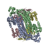


 Mass: 290.273 Da / Num. of mol.: 3 / Source method: obtained synthetically / Formula: C10H18N4O6
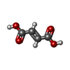
 Mass: 116.072 Da / Num. of mol.: 1 / Source method: obtained synthetically / Formula: C4H4O4
 Mass: 62.068 Da / Num. of mol.: 5 / Source method: obtained synthetically / Formula: C2H6O2
 Mass: 106.120 Da / Num. of mol.: 1 / Source method: obtained synthetically / Formula: C4H10O3
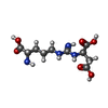
 Type: L-peptide linking / Mass: 175.209 Da / Num. of mol.: 1 / Source method: obtained synthetically / Formula: C6H15N4O2
 Sample preparation
 Processing