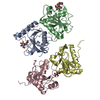
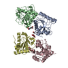
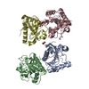
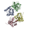
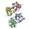
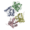
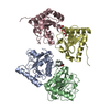
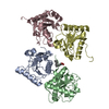
[English] 日本語
Yorodumi
- PDB-6idm: Crystal structure of Peptidoglycan recognition protein (PGRP-S) w... -

+
Open data


ID or keywords:

Loading...

-
Basic information

Entry
Database: PDB / ID: 6idm
TitleCrystal structure of Peptidoglycan recognition protein (PGRP-S) with Tartaric acid at 3.20 A resolution
ComponentsPeptidoglycan recognition protein 1
KeywordsIMMUNE SYSTEM
Function / homology
Function and homology information


peptidoglycan immune receptor activity / N-acetylmuramoyl-L-alanine amidase activity / peptidoglycan binding / negative regulation of cytokine production / detection of bacterium / peptidoglycan catabolic process / defense response to Gram-positive bacterium / innate immune response / zinc ion binding / extracellular region
Similarity search - Function
Peptidoglycan recognition protein, PGRP-S / Peptidoglycan recognition protein family domain, metazoa/bacteria / Peptidoglycan recognition protein / Animal peptidoglycan recognition proteins homologous to Bacteriophage T3 lysozyme. / Lysozyme-like / Peptidoglycan recognition protein-like / Ami_2 / N-acetylmuramoyl-L-alanine amidase / N-acetylmuramoyl-L-alanine amidase domain / N-acetylmuramoyl-L-alanine amidase/PGRP domain superfamily ...Peptidoglycan recognition protein, PGRP-S / Peptidoglycan recognition protein family domain, metazoa/bacteria / Peptidoglycan recognition protein / Animal peptidoglycan recognition proteins homologous to Bacteriophage T3 lysozyme. / Lysozyme-like / Peptidoglycan recognition protein-like / Ami_2 / N-acetylmuramoyl-L-alanine amidase / N-acetylmuramoyl-L-alanine amidase domain / N-acetylmuramoyl-L-alanine amidase/PGRP domain superfamily / 3-Layer(aba) Sandwich / Alpha Beta
Similarity search - Domain/homology
L(+)-TARTARIC ACID / Peptidoglycan recognition protein 1
Similarity search - Component
Biological speciesCamelus dromedarius (Arabian camel)
MethodX-RAY DIFFRACTION / SYNCHROTRON / MOLECULAR REPLACEMENT / Resolution: 3.2 Å
AuthorsBairagya, H.R. / Shokeen, A. / Sharma, P. / Singh, P.K. / Sharma, S. / Singh, T.P.
CitationJournal: To Be Published
Title: Crystal structure of Peptidoglycan recognition protein (PGRP-S) with Tartaric acid at 3.20 A resolution
Authors: Bairagya, H.R. / Shokeen, A. / Sharma, P. / Singh, P.K. / Sharma, S. / Singh, T.P.
History
DepositionSep 10, 2018Deposition site: PDBJ / Processing site: PDBJ
Revision 1.0Sep 26, 2018Provider: repository / Type: Initial release
Revision 1.1Nov 22, 2023Group: Data collection / Database references / Refinement description
Category: chem_comp_atom / chem_comp_bond ...chem_comp_atom / chem_comp_bond / database_2 / pdbx_initial_refinement_model
Item: _database_2.pdbx_DOI / _database_2.pdbx_database_accession

-
Structure visualization

Structure viewerMolecule:
MolmilJmol/JSmol

Downloads & links

-
Assembly

Deposited unit
A: Peptidoglycan recognition protein 1
B: Peptidoglycan recognition protein 1
C: Peptidoglycan recognition protein 1
D: Peptidoglycan recognition protein 1
hetero molecules


Theoretical massNumber of molelcules
Total (without water)74,1585
Polymers74,0084
Non-polymers1501
Water2,234124
1


  • Idetical with deposited unit
  • defined by author
  • Evidence: Tetramer from crystallographic data
TypeNameSymmetry operationNumber
identity operation1_555x,y,z1
Unit cell
Length a, b, c (Å)88.309, 101.149, 163.566
Angle α, β, γ (deg.)90.00, 90.00, 90.00
Int Tables number23
Space group name H-MI222

-
Components

#1: Protein
Peptidoglycan recognition protein 1 / Peptidoglycan recognition protein short / PGRP-S


Mass: 18501.947 Da / Num. of mol.: 4 / Source method: isolated from a natural source / Source: (natural) Camelus dromedarius (Arabian camel) / References: UniProt: Q9GK12
#2: Chemical ChemComp-TLA / L(+)-TARTARIC ACID


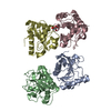
Mass: 150.087 Da / Num. of mol.: 1 / Source method: obtained synthetically / Formula: C4H6O6
#3: Water ChemComp-HOH / water


Mass: 18.015 Da / Num. of mol.: 124 / Source method: isolated from a natural source / Formula: H2O

-
Experimental details

-
Experiment

ExperimentMethod: X-RAY DIFFRACTION / Number of used crystals: 1

-
Sample preparation

CrystalDensity Matthews: 2.47 Å3/Da / Density % sol: 50.15 %
Crystal growTemperature: 298 K / Method: vapor diffusion, hanging drop / pH: 8 / Details: 12% PEG 3350, 100mM Sodium potassium tartarate

-
Data collection

DiffractionMean temperature: 298 K
Diffraction sourceSource: SYNCHROTRON / Site: ESRF / Beamline: BM14 / Wavelength: 0.9537 Å
DetectorType: DECTRIS PILATUS3 6M / Detector: PIXEL / Date: May 13, 2018
RadiationProtocol: SINGLE WAVELENGTH / Monochromatic (M) / Laue (L): M / Scattering type: x-ray
Radiation wavelengthWavelength: 0.9537 Å / Relative weight: 1
ReflectionResolution: 3.2→47.99 Å / Num. obs: 12435 / % possible obs: 100 % / Redundancy: 6.88 % / Rsym value: 0.27 / Net I/σ(I): 4.27
Reflection shellResolution: 3.2→3.28 Å / Rmerge(I) obs: 0.63 / Num. unique obs: 853 / Rrim(I) all: 0.35 / % possible all: 100

-
Processing

Software
NameVersionClassification
REFMAC5.8.0158refinement
HKL-2000data reduction
SCALEPACKdata scaling
PHASERphasing
RefinementMethod to determine structure: MOLECULAR REPLACEMENT
Starting model: 3RT4
Resolution: 3.2→47.99 Å / Cor.coef. Fo:Fc: 0.953 / Cor.coef. Fo:Fc free: 0.89 / SU B: 38.738 / SU ML: 0.596 / Cross valid method: THROUGHOUT / ESU R Free: 0.574 / Details: HYDROGENS HAVE BEEN ADDED IN THE RIDING POSITIONS
RfactorNum. reflection% reflectionSelection details
Rfree0.2654 458 3.7 %RANDOM
Rwork0.19193 ---
obs0.19478 11977 99.94 %-
Solvent computationIon probe radii: 0.8 Å / Shrinkage radii: 0.8 Å / VDW probe radii: 1.2 Å
Displacement parametersBiso mean: 75.203 Å2
Baniso -1Baniso -2Baniso -3
1-15.73 Å20 Å2-0 Å2
2---10.48 Å20 Å2
3----5.25 Å2
Refinement stepCycle: 1 / Resolution: 3.2→47.99 Å
ProteinNucleic acidLigandSolventTotal
Num. atoms5200 0 10 124 5334
Refine LS restraints
Refine-IDTypeDev idealDev ideal targetNumber
X-RAY DIFFRACTIONr_bond_refined_d0.0090.0195361
X-RAY DIFFRACTIONr_bond_other_d0.0020.024786
X-RAY DIFFRACTIONr_angle_refined_deg1.41.9237296
X-RAY DIFFRACTIONr_angle_other_deg2.077311026
X-RAY DIFFRACTIONr_dihedral_angle_1_deg7.0545660
X-RAY DIFFRACTIONr_dihedral_angle_2_deg38.4621.846260
X-RAY DIFFRACTIONr_dihedral_angle_3_deg15.25115804
X-RAY DIFFRACTIONr_dihedral_angle_4_deg13.7651564
X-RAY DIFFRACTIONr_chiral_restr0.080.2758
X-RAY DIFFRACTIONr_gen_planes_refined0.0050.0216060
X-RAY DIFFRACTIONr_gen_planes_other0.0020.021212
X-RAY DIFFRACTIONr_nbd_refined
X-RAY DIFFRACTIONr_nbd_other
X-RAY DIFFRACTIONr_nbtor_refined
X-RAY DIFFRACTIONr_nbtor_other
X-RAY DIFFRACTIONr_xyhbond_nbd_refined
X-RAY DIFFRACTIONr_xyhbond_nbd_other
X-RAY DIFFRACTIONr_metal_ion_refined
X-RAY DIFFRACTIONr_metal_ion_other
X-RAY DIFFRACTIONr_symmetry_vdw_refined
X-RAY DIFFRACTIONr_symmetry_vdw_other
X-RAY DIFFRACTIONr_symmetry_hbond_refined
X-RAY DIFFRACTIONr_symmetry_hbond_other
X-RAY DIFFRACTIONr_symmetry_metal_ion_refined
X-RAY DIFFRACTIONr_symmetry_metal_ion_other
X-RAY DIFFRACTIONr_mcbond_it10.1887.6292652
X-RAY DIFFRACTIONr_mcbond_other10.197.6292651
X-RAY DIFFRACTIONr_mcangle_it16.33711.4343308
X-RAY DIFFRACTIONr_mcangle_other16.33511.4343309
X-RAY DIFFRACTIONr_scbond_it9.8647.8082709
X-RAY DIFFRACTIONr_scbond_other9.8697.8082706
X-RAY DIFFRACTIONr_scangle_it
X-RAY DIFFRACTIONr_scangle_other10.37711.6443987
X-RAY DIFFRACTIONr_long_range_B_refined14.49288.1486041
X-RAY DIFFRACTIONr_long_range_B_other14.47488.1536030
X-RAY DIFFRACTIONr_rigid_bond_restr
X-RAY DIFFRACTIONr_sphericity_free
X-RAY DIFFRACTIONr_sphericity_bonded
LS refinement shellResolution: 3.2→3.283 Å / Total num. of bins used: 20
RfactorNum. reflection% reflection
Rfree0.368 33 -
Rwork0.34 853 -
obs--100 %

+
About Yorodumi

-
News

-
Feb 9, 2022. New format data for meta-information of EMDB entries

New format data for meta-information of EMDB entries

  • Version 3 of the EMDB header file is now the official format.
  • The previous official version 1.9 will be removed from the archive.

Related info.:EMDB header

External links:wwPDB to switch to version 3 of the EMDB data model

-
Aug 12, 2020. Covid-19 info

Covid-19 info

URL: https://pdbjlvh1.pdbj.org/emnavi/covid19.php

New page: Covid-19 featured information page in EM Navigator.

Related info.:Covid-19 info / Mar 5, 2020. Novel coronavirus structure data

+
Mar 5, 2020. Novel coronavirus structure data

Novel coronavirus structure data

Related info.:Yorodumi Speices / Aug 12, 2020. Covid-19 info

External links:COVID-19 featured content - PDBj / Molecule of the Month (242):Coronavirus Proteases

+
Jan 31, 2019. EMDB accession codes are about to change! (news from PDBe EMDB page)

EMDB accession codes are about to change! (news from PDBe EMDB page)

  • The allocation of 4 digits for EMDB accession codes will soon come to an end. Whilst these codes will remain in use, new EMDB accession codes will include an additional digit and will expand incrementally as the available range of codes is exhausted. The current 4-digit format prefixed with “EMD-” (i.e. EMD-XXXX) will advance to a 5-digit format (i.e. EMD-XXXXX), and so on. It is currently estimated that the 4-digit codes will be depleted around Spring 2019, at which point the 5-digit format will come into force.
  • The EM Navigator/Yorodumi systems omit the EMD- prefix.

Related info.:Q: What is EMD? / ID/Accession-code notation in Yorodumi/EM Navigator

External links:EMDB Accession Codes are Changing Soon! / Contact to PDBj

+
Jul 12, 2017. Major update of PDB

Major update of PDB

  • wwPDB released updated PDB data conforming to the new PDBx/mmCIF dictionary.
  • This is a major update changing the version number from 4 to 5, and with Remediation, in which all the entries are updated.
  • In this update, many items about electron microscopy experimental information are reorganized (e.g. em_software).
  • Now, EM Navigator and Yorodumi are based on the updated data.

External links:wwPDB Remediation / Enriched Model Files Conforming to OneDep Data Standards Now Available in the PDB FTP Archive

-
Yorodumi

Thousand views of thousand structures

  • Yorodumi is a browser for structure data from EMDB, PDB, SASBDB, etc.
  • This page is also the successor to EM Navigator detail page, and also detail information page/front-end page for Omokage search.
  • The word "yorodu" (or yorozu) is an old Japanese word meaning "ten thousand". "mi" (miru) is to see.

Related info.:EMDB / PDB / SASBDB / Comparison of 3 databanks / Yorodumi Search / Aug 31, 2016. New EM Navigator & Yorodumi / Yorodumi Papers / Jmol/JSmol / Function and homology information / Changes in new EM Navigator and Yorodumi

Read more