 Movie
Movie Controller

[English] 日本語
 Yorodumi
- PDB-3cxa: Crystal structure of the complex of peptidoglycan recognition pro... -
+ Open data

- Basic information
| Entry | Database: PDB / ID: 3cxa | |||||||||
|---|---|---|---|---|---|---|---|---|---|---|
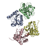
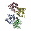
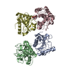
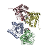
| Title | Crystal structure of the complex of peptidoglycan recognition protein with alpha-D-glucopyranosyl alpha-D-glucopyranoside at 3.4 A resolution | |||||||||
 Components Components | Peptidoglycan recognition protein | |||||||||
 Keywords Keywords | ANTIBIOTIC / Peptidoglycan recognition protein / complex / trehalose / immune response / secreted / antimicrobial / PGRP | |||||||||
| Function / homology |  Function and homology information Function and homology informationpeptidoglycan immune receptor activity / N-acetylmuramoyl-L-alanine amidase activity / peptidoglycan binding / negative regulation of cytokine production / detection of bacterium / peptidoglycan catabolic process / defense response to Gram-positive bacterium / innate immune response / extracellular region / zinc ion binding Similarity search - Function | |||||||||
| Biological species |  | |||||||||
| Method |  X-RAY DIFFRACTION / MOLECULAR REPLACEMENT / Resolution: 3.4 Å X-RAY DIFFRACTION / MOLECULAR REPLACEMENT / Resolution: 3.4 Å | |||||||||
 Authors Authors | Balaji, K. / Sharma, P. / Singh, N. / Sinha, M. / Bhushan, A. / Kaur, P. / Sharma, S. / Singh, T.P. | |||||||||
 Citation Citation | Journal: To be Published Title: Crystal structure of the complex of peptidoglycan recognition protein with alpha-D-glucopyranosyl alpha-D-glucopyranoside at 3.4 A resolution Authors: Balaji, K. / Sharma, P. / Singh, N. / Sinha, M. / Bhushan, A. / Kaur, P. / Sharma, S. / Singh, T.P. | |||||||||
| History |
|
- Structure visualization
| Structure viewer | Molecule: MolmilJmol/JSmol |
|---|
- Downloads & links
-Download
| PDBx/mmCIF format | 3cxa.cif.gz | 140.2 KB | Display | PDBx/mmCIF format |
|---|---|---|---|---|
| PDB format | pdb3cxa.ent.gz | 112.1 KB | Display | PDB format |
| PDBx/mmJSON format | 3cxa.json.gz | Tree view | PDBx/mmJSON format | |
| Others |  Other downloads Other downloads |
-Validation report
| Arichive directory | https://data.pdbj.org/pub/pdb/validation_reports/cx/3cxaftp://data.pdbj.org/pub/pdb/validation_reports/cx/3cxa | HTTPS FTP |
|---|
-Related structure data
| Related structure data |  2r2kS  3c93 S: Starting model for refinement |
|---|---|
| Similar structure data |


-Links
 PDBj
PDBj

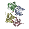
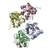
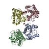
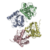
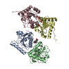
- Assembly
| Deposited unit | 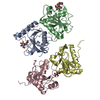
| ||||||||
|---|---|---|---|---|---|---|---|---|---|
| 1 |
| ||||||||
| 2 | 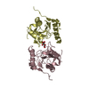
| ||||||||
| 3 | 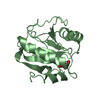
| ||||||||
| 4 | 
| ||||||||
| Unit cell |
|
-Components
| #1: Protein | Mass: 19011.459 Da / Num. of mol.: 4 / Source method: isolated from a natural source / Source: (natural) #2: Polysaccharide | 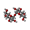  Source method: isolated from a genetically manipulated source Details: oligosaccharide with reducing-end-to-reducing-end glycosidic bond References: trehalose #3: Chemical | ChemComp-TLA / |   Mass: 150.087 Da / Num. of mol.: 1 / Source method: obtained synthetically / Formula: C4H6O6 Mass: 150.087 Da / Num. of mol.: 1 / Source method: obtained synthetically / Formula: C4H6O6Has protein modification | Y | |
|---|
-Experimental details
-Experiment
| Experiment | Method: X-RAY DIFFRACTION / Number of used crystals: 1 |
|---|
- Sample preparation
| Crystal | Density Matthews: 2.47 Å3/Da / Density % sol: 50.21 % |
|---|---|
| Crystal grow | Temperature: 298 K / Method: vapor diffusion, hanging drop / pH: 7.2 Details: 0.2M SODIUM POTASSIUM TARTRATE, 10% PEG 3350, pH 7.2, VAPOR DIFFUSION, HANGING DROP, temperature 298K |
-Data collection
| Diffraction | Mean temperature: 300 K |
|---|---|
| Diffraction source | Source: ROTATING ANODE / Type: RIGAKU RU300 / Wavelength: 1.5418 Å |
| Detector | Type: MARRESEARCH / Detector: IMAGE PLATE / Date: Nov 22, 2007 / Details: MIRROR |
| Radiation | Monochromator: GRAPHITE / Protocol: SINGLE WAVELENGTH / Monochromatic (M) / Laue (L): M / Scattering type: x-ray |
| Radiation wavelength | Wavelength: 1.5418 Å / Relative weight: 1 |
| Reflection | Resolution: 3.4→20 Å / Num. obs: 10724 / % possible obs: 98.3 % / Observed criterion σ(F): 0 / Observed criterion σ(I): 0 / Biso Wilson estimate: 39.5 Å2 / Rsym value: 0.182 / Net I/σ(I): 4.4 |
| Reflection shell | Resolution: 3.4→3.52 Å / Mean I/σ(I) obs: 1.9 / Rsym value: 0.545 / % possible all: 98.9 |
- Processing
| Software |
| |||||||||||||||||||||||||
|---|---|---|---|---|---|---|---|---|---|---|---|---|---|---|---|---|---|---|---|---|---|---|---|---|---|---|
| Refinement | Method to determine structure: MOLECULAR REPLACEMENT Starting model: 2R2K Resolution: 3.4→20 Å / Cross valid method: THROUGHOUT / σ(F): 0 / σ(I): 0 / Stereochemistry target values: MAXIMUM LIKELIHOOD
| |||||||||||||||||||||||||
| Refinement step | Cycle: LAST / Resolution: 3.4→20 Å
| |||||||||||||||||||||||||
| Refine LS restraints |
|
