 ムービー
ムービー コントローラー


+ データを開く

- 基本情報
基本情報
| 登録情報 | データベース: PDB / ID: 7dy5 | |||||||||
|---|---|---|---|---|---|---|---|---|---|---|
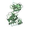
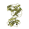
| タイトル | Structure of the ternary complex of peptidoglycan recognition protein-short (PGRP-S) with hexanoic acid and tartaric acid at 2.30A resolution | |||||||||
 要素 要素 | Peptidoglycan recognition protein 1 | |||||||||
 キーワード キーワード | IMMUNE SYSTEM / Recognition protein | |||||||||
| 機能・相同性 |  機能・相同性情報 機能・相同性情報peptidoglycan immune receptor activity / N-acetylmuramoyl-L-alanine amidase activity / peptidoglycan binding / negative regulation of cytokine production / detection of bacterium / peptidoglycan catabolic process / defense response to Gram-positive bacterium / innate immune response / extracellular region / zinc ion binding 類似検索 - 分子機能 | |||||||||
| 生物種 |  | |||||||||
| 手法 |  X線回折 / シンクロトロン / 分子置換 / 解像度: 2.3 Å X線回折 / シンクロトロン / 分子置換 / 解像度: 2.3 Å | |||||||||
 データ登録者 データ登録者 | Maurya, A. / Viswanathan, V. / Sharma, P. / Sharma, S. / Singh, T.P. | |||||||||
 引用 引用 | ジャーナル: To Be Published タイトル: Structure of the ternary complex of peptidoglycan recognition protein-short (PGRP-S) 著者: Maurya, A. / Viswanathan, V. / Sharma, P. / Sharma, S. / Singh, T.P. | |||||||||
| 履歴 |
|
- 構造の表示
構造の表示
| 構造ビューア | 分子: MolmilJmol/JSmol |
|---|
- ダウンロードとリンク
-ダウンロード
| PDBx/mmCIF形式 | 7dy5.cif.gz | 182.3 KB | 表示 | PDBx/mmCIF形式 |
|---|---|---|---|---|
| PDB形式 | pdb7dy5.ent.gz | 118.2 KB | 表示 | PDB形式 |
| PDBx/mmJSON形式 | 7dy5.json.gz | ツリー表示 | PDBx/mmJSON形式 | |
| その他 |  その他のダウンロード その他のダウンロード |
-検証レポート
| アーカイブディレクトリ | https://data.pdbj.org/pub/pdb/validation_reports/dy/7dy5ftp://data.pdbj.org/pub/pdb/validation_reports/dy/7dy5 | HTTPS FTP |
|---|
-関連構造データ
| 関連構造データ |  6j3w S: 精密化の開始モデル |
|---|---|
| 類似構造データ |


-リンク
 PDBj
PDBj
- 集合体
集合体
| 登録構造単位 | 
| |||||||||||||||
|---|---|---|---|---|---|---|---|---|---|---|---|---|---|---|---|---|
| 1 | 
| |||||||||||||||
| 2 | 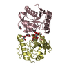
| |||||||||||||||
| 単位格子 |
| |||||||||||||||
| Components on special symmetry positions |
|
-要素
-タンパク質 , 1種, 4分子 ABCD
| #1: タンパク質 | 分子量: 19011.459 Da / 分子数: 4 / 由来タイプ: 天然 由来: (天然) 参照: UniProt: Q9GK12 |
|---|
-非ポリマー , 6種, 433分子 


| #2: 化合物 |  分子量: 150.087 Da / 分子数: 3 / 由来タイプ: 合成 / 式: C4H6O6 / タイプ: SUBJECT OF INVESTIGATION 分子量: 150.087 Da / 分子数: 3 / 由来タイプ: 合成 / 式: C4H6O6 / タイプ: SUBJECT OF INVESTIGATION#3: 化合物 | ChemComp-EDO /  分子量: 62.068 Da / 分子数: 4 / 由来タイプ: 合成 / 式: C2H6O2 / タイプ: SUBJECT OF INVESTIGATION 分子量: 62.068 Da / 分子数: 4 / 由来タイプ: 合成 / 式: C2H6O2 / タイプ: SUBJECT OF INVESTIGATION#4: 化合物 | ChemComp-CL / |  分子量: 35.453 Da / 分子数: 1 / 由来タイプ: 合成 / 式: Cl / タイプ: SUBJECT OF INVESTIGATION 分子量: 35.453 Da / 分子数: 1 / 由来タイプ: 合成 / 式: Cl / タイプ: SUBJECT OF INVESTIGATION#5: 化合物 | ChemComp-6NA / |  分子量: 116.158 Da / 分子数: 1 / 由来タイプ: 合成 / 式: C6H12O2 / タイプ: SUBJECT OF INVESTIGATION 分子量: 116.158 Da / 分子数: 1 / 由来タイプ: 合成 / 式: C6H12O2 / タイプ: SUBJECT OF INVESTIGATION#6: 化合物 |  分子量: 92.094 Da / 分子数: 2 / 由来タイプ: 合成 / 式: C3H8O3 / タイプ: SUBJECT OF INVESTIGATION 分子量: 92.094 Da / 分子数: 2 / 由来タイプ: 合成 / 式: C3H8O3 / タイプ: SUBJECT OF INVESTIGATION#7: 水 | ChemComp-HOH / | 分子量: 18.015 Da / 分子数: 422 / 由来タイプ: 天然 / 式: H2O |
|---|
-詳細
| 研究の焦点であるリガンドがあるか | Y |
|---|---|
| Has protein modification | Y |
-実験情報
-実験
| 実験 | 手法: X線回折 / 使用した結晶の数: 1 |
|---|
- 試料調製
試料調製
| 結晶 | マシュー密度: 2.39 Å3/Da / 溶媒含有率: 48.55 % |
|---|---|
| 結晶化 | 温度: 298 K / 手法: 蒸気拡散法, ハンギングドロップ法 / pH: 8.5 詳細: 10% PEG 3350, 0.2M Sodium potassium tartrate, 20% Glycerol |
-データ収集
| 回折 | 平均測定温度: 100 K / Serial crystal experiment: N |
|---|---|
| 放射光源 | 由来: シンクロトロン / サイト: ESRF  / ビームライン: ID30B / 波長: 0.97 Å / ビームライン: ID30B / 波長: 0.97 Å |
| 検出器 | タイプ: DECTRIS PILATUS3 2M / 検出器: PIXEL / 日付: 2018年11月18日 |
| 放射 | モノクロメーター: M / プロトコル: SINGLE WAVELENGTH / 単色(M)・ラウエ(L): M / 散乱光タイプ: x-ray |
| 放射波長 | 波長: 0.97 Å / 相対比: 1 |
| 反射 | 解像度: 2.3→50.68 Å / Num. obs: 25883 / % possible obs: 78.01 % / 冗長度: 4.8 % / Biso Wilson estimate: 23.44 Å2 / CC1/2: 0.99 / Rmerge(I) obs: 0.11 / Rpim(I) all: 0.05 / Net I/σ(I): 8.1 |
| 反射 シェル | 解像度: 2.3→2.39 Å / 冗長度: 1.8 % / Rmerge(I) obs: 0.64 / Mean I/σ(I) obs: 2.4 / Num. unique obs: 1879 / CC1/2: 0.87 / Rpim(I) all: 0.32 / % possible all: 58 |
- 解析
解析
| ソフトウェア |
| ||||||||||||||||||||||||||||||||||||||||||||||||||||||||||||||||||||||
|---|---|---|---|---|---|---|---|---|---|---|---|---|---|---|---|---|---|---|---|---|---|---|---|---|---|---|---|---|---|---|---|---|---|---|---|---|---|---|---|---|---|---|---|---|---|---|---|---|---|---|---|---|---|---|---|---|---|---|---|---|---|---|---|---|---|---|---|---|---|---|---|
| 精密化 | 構造決定の手法: 分子置換 開始モデル: 6J3W 6j3w 解像度: 2.3→50.68 Å / SU ML: 0.2923 / 交差検証法: FREE R-VALUE / σ(F): 1.38 / 位相誤差: 24.0535 立体化学のターゲット値: GeoStd + Monomer Library + CDL v1.2
| ||||||||||||||||||||||||||||||||||||||||||||||||||||||||||||||||||||||
| 溶媒の処理 | 減衰半径: 0.9 Å / VDWプローブ半径: 1.11 Å / 溶媒モデル: FLAT BULK SOLVENT MODEL | ||||||||||||||||||||||||||||||||||||||||||||||||||||||||||||||||||||||
| 原子変位パラメータ | Biso mean: 26.25 Å2 | ||||||||||||||||||||||||||||||||||||||||||||||||||||||||||||||||||||||
| 精密化ステップ | サイクル: LAST / 解像度: 2.3→50.68 Å
| ||||||||||||||||||||||||||||||||||||||||||||||||||||||||||||||||||||||
| 拘束条件 |
| ||||||||||||||||||||||||||||||||||||||||||||||||||||||||||||||||||||||
| LS精密化 シェル |
|
