 Movie
Movie Controller

[English] 日本語
 Yorodumi
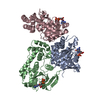
- PDB-6i8h: Structure of the plant immune signaling node EDS1 (enhanced disea... -
+ Open data

- Basic information
| Entry | Database: PDB / ID: 6i8h | ||||||
|---|---|---|---|---|---|---|---|
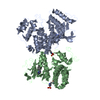
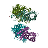
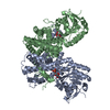
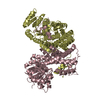
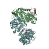
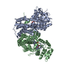
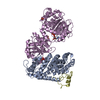
| Title | Structure of the plant immune signaling node EDS1 (enhanced disease susceptibility 1) in complex with nanobody ENB15 | ||||||
 Components Components |
| ||||||
 Keywords Keywords | IMMUNE SYSTEM / enhanced disease susceptibility 1 / plant innate immune system / alpha/beta hydrolase fold / nanobody | ||||||
| Function / homology |  Function and homology information Function and homology informationlipid metabolic process / defense response / hydrolase activity / nucleus / cytoplasm Similarity search - Function | ||||||
| Biological species |   | ||||||
| Method |  X-RAY DIFFRACTION / SYNCHROTRON / MOLECULAR REPLACEMENT / Resolution: 3.682 Å X-RAY DIFFRACTION / SYNCHROTRON / MOLECULAR REPLACEMENT / Resolution: 3.682 Å | ||||||
 Authors Authors | Niefind, K. / Voss, M. / Toelzer, C. | ||||||
| Funding support |  Germany, 1items Germany, 1items
| ||||||
 Citation Citation | Journal: J.Struct.Biol. / Year: 2019 Title: Arabidopsis immunity regulator EDS1 in a PAD4/SAG101-unbound form is a monomer with an inherently inactive conformation. Authors: Voss, M. / Toelzer, C. / Bhandari, D.D. / Parker, J.E. / Niefind, K. #1: Journal: Cell Host Microbe / Year: 2013Title: Structural basis for signaling by exclusive EDS1 heteromeric complexes with SAG101 or PAD4 in plant innate immunity. Authors: Wagner, S. / Stuttmann, J. / Rietz, S. / Guerois, R. / Brunstein, E. / Bautor, J. / Niefind, K. / Parker, J.E. #2: Journal: Acta Crystallogr. Sect. F Struct. Biol. Cryst. Commun. Year: 2011 Title: Crystallization and preliminary crystallographic analysis of Arabidopsis thaliana EDS1, a key component of plant immunity, in complex with its signalling partner SAG101. Authors: Wagner, S. / Rietz, S. / Parker, J.E. / Niefind, K. #3: Journal: New Phytol. / Year: 2011 Title: Different roles of Enhanced Disease Susceptibility1 (EDS1) bound to and dissociated from Phytoalexin Deficient4 (PAD4) in Arabidopsis immunity. Authors: Rietz, S. / Stamm, A. / Malonek, S. / Wagner, S. / Becker, D. / Medina-Escobar, N. / Vlot, A.C. / Feys, B.J. / Niefind, K. / Parker, J.E. | ||||||
| History |
|
- Structure visualization
| Structure viewer | Molecule: MolmilJmol/JSmol |
|---|
- Downloads & links
-Download
| PDBx/mmCIF format | 6i8h.cif.gz | 317.5 KB | Display | PDBx/mmCIF format |
|---|---|---|---|---|
| PDB format | pdb6i8h.ent.gz | 262.3 KB | Display | PDB format |
| PDBx/mmJSON format | 6i8h.json.gz | Tree view | PDBx/mmJSON format | |
| Others |  Other downloads Other downloads |
-Validation report
| Arichive directory | https://data.pdbj.org/pub/pdb/validation_reports/i8/6i8hftp://data.pdbj.org/pub/pdb/validation_reports/i8/6i8h | HTTPS FTP |
|---|
-Related structure data
| Related structure data |  6i8gC  6q6zC  4nfuS S: Starting model for refinement C: citing same article ( |
|---|---|
| Similar structure data |

-Links
 PDBj
PDBj


- Assembly
| Deposited unit | 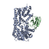
| ||||||||
|---|---|---|---|---|---|---|---|---|---|
| 1 |
| ||||||||
| Unit cell |
|
-Components
| #1: Protein | Mass: 72736.234 Da / Num. of mol.: 1 Source method: isolated from a genetically manipulated source Source: (gene. exp.)  |
|---|---|
| #2: Antibody | Mass: 14975.403 Da / Num. of mol.: 1 Source method: isolated from a genetically manipulated source Source: (gene. exp.) |
| Has protein modification | Y |
-Experimental details
-Experiment
| Experiment | Method: X-RAY DIFFRACTION / Number of used crystals: 1 |
|---|
- Sample preparation
| Crystal | Density Matthews: 2.66 Å3/Da / Density % sol: 53.8 % |
|---|---|
| Crystal grow | Temperature: 277.15 K / Method: vapor diffusion, sitting drop / pH: 6.5 Details: Reservoir composition: 12.5 % (v/v) (RS)-2-methyl-2,4-pentanediol, 12.5 % (w/v) PEG 1000, 12.5 % (w/v) PEG3350, 0.03 M magnesium chloride, 0.03 M calcium chloride, 0.0612 M MES, 0.0388 M ...Details: Reservoir composition: 12.5 % (v/v) (RS)-2-methyl-2,4-pentanediol, 12.5 % (w/v) PEG 1000, 12.5 % (w/v) PEG3350, 0.03 M magnesium chloride, 0.03 M calcium chloride, 0.0612 M MES, 0.0388 M imidazole, pH 6.5; protein stock solution: 4.1 mg/ml protein, 50 mM sodium chloride, 1 % (v/v) glycerole, 1 mM DTT, 50 mM HEPES, pH 8.0; drop composition: 150 nl protein stock solution plus 225 nl reservoir solution; cryo conditions: the crystals were flash frozen directly from the equilibrated crystallization drops. |
-Data collection
| Diffraction | Mean temperature: 100 K / Serial crystal experiment: N |
|---|---|
| Diffraction source | Source: SYNCHROTRON / Site: PETRA III, EMBL c/o DESY / Beamline: P13 (MX1) / Wavelength: 0.9763 Å |
| Detector | Type: DECTRIS PILATUS 6M / Detector: PIXEL / Date: Nov 12, 2016 |
| Radiation | Protocol: SINGLE WAVELENGTH / Monochromatic (M) / Laue (L): M / Scattering type: x-ray |
| Radiation wavelength | Wavelength: 0.9763 Å / Relative weight: 1 |
| Reflection | Resolution: 3.682→97.361 Å / Num. obs: 8246 / % possible obs: 93 % / Redundancy: 6.3 % / Rmerge(I) obs: 0.111 / Rsym value: 0.111 / Net I/σ(I): 9.8 |
| Reflection shell | Resolution: 3.682→4.05 Å / Redundancy: 7.4 % / Rmerge(I) obs: 1.349 / Mean I/σ(I) obs: 1.5 / Num. unique obs: 411 / Rsym value: 1.349 / % possible all: 59.5 |
- Processing
| Software |
| |||||||||||||||||||||||||||||||||||||||||||||||||||||||||||||||||||||||||||||||||||||||||||||||||||||||||||||||||||||||||||||||||||||||||||||||||||||||||||||||||||||||||||||||||||||||||||||||||||||||||||||||||||||||||||||||||||||||||||||||||||||||||||||||||||||||||||||||||||||||||||||||||||||||||||||||||||||||||||||||||||||
|---|---|---|---|---|---|---|---|---|---|---|---|---|---|---|---|---|---|---|---|---|---|---|---|---|---|---|---|---|---|---|---|---|---|---|---|---|---|---|---|---|---|---|---|---|---|---|---|---|---|---|---|---|---|---|---|---|---|---|---|---|---|---|---|---|---|---|---|---|---|---|---|---|---|---|---|---|---|---|---|---|---|---|---|---|---|---|---|---|---|---|---|---|---|---|---|---|---|---|---|---|---|---|---|---|---|---|---|---|---|---|---|---|---|---|---|---|---|---|---|---|---|---|---|---|---|---|---|---|---|---|---|---|---|---|---|---|---|---|---|---|---|---|---|---|---|---|---|---|---|---|---|---|---|---|---|---|---|---|---|---|---|---|---|---|---|---|---|---|---|---|---|---|---|---|---|---|---|---|---|---|---|---|---|---|---|---|---|---|---|---|---|---|---|---|---|---|---|---|---|---|---|---|---|---|---|---|---|---|---|---|---|---|---|---|---|---|---|---|---|---|---|---|---|---|---|---|---|---|---|---|---|---|---|---|---|---|---|---|---|---|---|---|---|---|---|---|---|---|---|---|---|---|---|---|---|---|---|---|---|---|---|---|---|---|---|---|---|---|---|---|---|---|---|---|---|---|---|---|---|---|---|---|---|---|---|---|---|---|---|---|---|---|---|---|---|---|---|---|---|---|---|---|---|---|---|---|---|---|---|---|---|---|---|---|---|---|---|---|---|---|---|---|---|---|---|---|
| Refinement | Method to determine structure: MOLECULAR REPLACEMENT Starting model: 4NFU Resolution: 3.682→72.94 Å / SU ML: 0.53 / Cross valid method: FREE R-VALUE / σ(F): 1.35 / Phase error: 35.75
| |||||||||||||||||||||||||||||||||||||||||||||||||||||||||||||||||||||||||||||||||||||||||||||||||||||||||||||||||||||||||||||||||||||||||||||||||||||||||||||||||||||||||||||||||||||||||||||||||||||||||||||||||||||||||||||||||||||||||||||||||||||||||||||||||||||||||||||||||||||||||||||||||||||||||||||||||||||||||||||||||||||
| Solvent computation | Shrinkage radii: 0.9 Å / VDW probe radii: 1.11 Å | |||||||||||||||||||||||||||||||||||||||||||||||||||||||||||||||||||||||||||||||||||||||||||||||||||||||||||||||||||||||||||||||||||||||||||||||||||||||||||||||||||||||||||||||||||||||||||||||||||||||||||||||||||||||||||||||||||||||||||||||||||||||||||||||||||||||||||||||||||||||||||||||||||||||||||||||||||||||||||||||||||||
| Refinement step | Cycle: LAST / Resolution: 3.682→72.94 Å
| |||||||||||||||||||||||||||||||||||||||||||||||||||||||||||||||||||||||||||||||||||||||||||||||||||||||||||||||||||||||||||||||||||||||||||||||||||||||||||||||||||||||||||||||||||||||||||||||||||||||||||||||||||||||||||||||||||||||||||||||||||||||||||||||||||||||||||||||||||||||||||||||||||||||||||||||||||||||||||||||||||||
| Refine LS restraints |
| |||||||||||||||||||||||||||||||||||||||||||||||||||||||||||||||||||||||||||||||||||||||||||||||||||||||||||||||||||||||||||||||||||||||||||||||||||||||||||||||||||||||||||||||||||||||||||||||||||||||||||||||||||||||||||||||||||||||||||||||||||||||||||||||||||||||||||||||||||||||||||||||||||||||||||||||||||||||||||||||||||||
| LS refinement shell |
| |||||||||||||||||||||||||||||||||||||||||||||||||||||||||||||||||||||||||||||||||||||||||||||||||||||||||||||||||||||||||||||||||||||||||||||||||||||||||||||||||||||||||||||||||||||||||||||||||||||||||||||||||||||||||||||||||||||||||||||||||||||||||||||||||||||||||||||||||||||||||||||||||||||||||||||||||||||||||||||||||||||
| Refinement TLS params. | Method: refined / Refine-ID: X-RAY DIFFRACTION
| |||||||||||||||||||||||||||||||||||||||||||||||||||||||||||||||||||||||||||||||||||||||||||||||||||||||||||||||||||||||||||||||||||||||||||||||||||||||||||||||||||||||||||||||||||||||||||||||||||||||||||||||||||||||||||||||||||||||||||||||||||||||||||||||||||||||||||||||||||||||||||||||||||||||||||||||||||||||||||||||||||||
| Refinement TLS group |
|
