 Movie
Movie Controller


+ Open data

- Basic information
| Entry | Database: PDB / ID: 6i2i | ||||||||||||
|---|---|---|---|---|---|---|---|---|---|---|---|---|---|
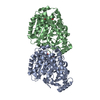
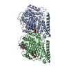
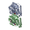
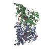
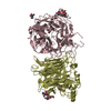
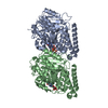
| Title | Refined 13pf Hela Cell Tubulin microtubule (EML4-NTD decorated) | ||||||||||||
 Components Components |
| ||||||||||||
 Keywords Keywords | STRUCTURAL PROTEIN / Microtubule / Tubulin / Hela / EML | ||||||||||||
| Function / homology |  Function and homology information Function and homology informationodontoblast differentiation / Post-chaperonin tubulin folding pathway / Cilium Assembly / cytoskeleton-dependent intracellular transport / Carboxyterminal post-translational modifications of tubulin / Microtubule-dependent trafficking of connexons from Golgi to the plasma membrane / Sealing of the nuclear envelope (NE) by ESCRT-III / Intraflagellar transport / Formation of tubulin folding intermediates by CCT/TriC / Gap junction assembly ...odontoblast differentiation / Post-chaperonin tubulin folding pathway / Cilium Assembly / cytoskeleton-dependent intracellular transport / Carboxyterminal post-translational modifications of tubulin / Microtubule-dependent trafficking of connexons from Golgi to the plasma membrane / Sealing of the nuclear envelope (NE) by ESCRT-III / Intraflagellar transport / Formation of tubulin folding intermediates by CCT/TriC / Gap junction assembly / Kinesins / Assembly and cell surface presentation of NMDA receptors / COPI-independent Golgi-to-ER retrograde traffic / GTPase activating protein binding / COPI-dependent Golgi-to-ER retrograde traffic / natural killer cell mediated cytotoxicity / nuclear envelope lumen / regulation of synapse organization / Recycling pathway of L1 / RHOH GTPase cycle / MHC class I protein binding / microtubule-based process / intercellular bridge / RHO GTPases activate IQGAPs / Hedgehog 'off' state / COPI-mediated anterograde transport / cytoplasmic microtubule / Activation of AMPK downstream of NMDARs / Loss of Nlp from mitotic centrosomes / Loss of proteins required for interphase microtubule organization from the centrosome / Recruitment of mitotic centrosome proteins and complexes / MHC class II antigen presentation / Recruitment of NuMA to mitotic centrosomes / cellular response to interleukin-4 / Anchoring of the basal body to the plasma membrane / HSP90 chaperone cycle for steroid hormone receptors (SHR) in the presence of ligand / Mitotic Prometaphase / EML4 and NUDC in mitotic spindle formation / AURKA Activation by TPX2 / ciliary tip / Resolution of Sister Chromatid Cohesion / Translocation of SLC2A4 (GLUT4) to the plasma membrane / sperm end piece / RHO GTPases Activate Formins / PKR-mediated signaling / structural constituent of cytoskeleton / microtubule cytoskeleton organization / HCMV Early Events / Aggrephagy / cytoplasmic ribonucleoprotein granule / azurophil granule lumen / mitotic spindle / The role of GTSE1 in G2/M progression after G2 checkpoint / Separation of Sister Chromatids / Regulation of PLK1 Activity at G2/M Transition / mitotic cell cycle / double-stranded RNA binding / sperm principal piece / microtubule cytoskeleton / cell body / Potential therapeutics for SARS / cytoskeleton / microtubule / Hydrolases; Acting on acid anhydrides; Acting on GTP to facilitate cellular and subcellular movement / cilium / membrane raft / protein domain specific binding / cell division / GTPase activity / Neutrophil degranulation / ubiquitin protein ligase binding / GTP binding / protein-containing complex binding / structural molecule activity / protein-containing complex / extracellular exosome / extracellular region / metal ion binding / nucleus / cytoplasm / cytosol Similarity search - Function | ||||||||||||
| Biological species |  Homo sapiens (human) Homo sapiens (human) | ||||||||||||
| Method | ELECTRON MICROSCOPY / single particle reconstruction / cryo EM / Resolution: 3.6 Å | ||||||||||||
 Authors Authors | Atherton, J.M. / Moores, C.A. | ||||||||||||
| Funding support |  United Kingdom, 3items United Kingdom, 3items
| ||||||||||||
 Citation Citation | Journal: Sci Signal / Year: 2019 Title: Mitotic phosphorylation by NEK6 and NEK7 reduces the microtubule affinity of EML4 to promote chromosome congression. Authors: Rozita Adib / Jessica M Montgomery / Joseph Atherton / Laura O'Regan / Mark W Richards / Kees R Straatman / Daniel Roth / Anne Straube / Richard Bayliss / Carolyn A Moores / Andrew M Fry / Abstract: EML4 is a microtubule-associated protein that promotes microtubule stability. We investigated its regulation across the cell cycle and found that EML4 was distributed as punctate foci along the ...EML4 is a microtubule-associated protein that promotes microtubule stability. We investigated its regulation across the cell cycle and found that EML4 was distributed as punctate foci along the microtubule lattice in interphase but exhibited reduced association with spindle microtubules in mitosis. Microtubule sedimentation and cryo-electron microscopy with 3D reconstruction revealed that the basic N-terminal domain of EML4 mediated its binding to the acidic C-terminal tails of α- and β-tubulin on the microtubule surface. The mitotic kinases NEK6 and NEK7 phosphorylated the EML4 N-terminal domain at Ser and Ser in vitro, and depletion of these kinases in cells led to increased EML4 binding to microtubules in mitosis. An S144A-S146A double mutant not only bound inappropriately to mitotic microtubules but also increased their stability and interfered with chromosome congression. In addition, constitutive activation of NEK6 or NEK7 reduced the association of EML4 with interphase microtubules. Together, these data support a model in which NEK6- and NEK7-dependent phosphorylation promotes the dissociation of EML4 from microtubules in mitosis in a manner that is required for efficient chromosome congression. | ||||||||||||
| History |
|
- Structure visualization
| Movie |
Movie viewer |
|---|---|
| Structure viewer | Molecule: MolmilJmol/JSmol |
- Downloads & links
-Download
| PDBx/mmCIF format | 6i2i.cif.gz | 166.6 KB | Display | PDBx/mmCIF format |
|---|---|---|---|---|
| PDB format | pdb6i2i.ent.gz | 127.7 KB | Display | PDB format |
| PDBx/mmJSON format | 6i2i.json.gz | Tree view | PDBx/mmJSON format | |
| Others |  Other downloads Other downloads |
-Validation report
| Arichive directory | https://data.pdbj.org/pub/pdb/validation_reports/i2/6i2iftp://data.pdbj.org/pub/pdb/validation_reports/i2/6i2i | HTTPS FTP |
|---|
-Related structure data
| Related structure data | 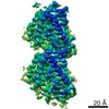 0331MC M: map data used to model this data C: citing same article ( |
|---|---|
| Similar structure data |


-Links
 PDBj
PDBj


- Assembly
| Deposited unit | 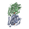
|
|---|---|
| 1 |
|
-Components
-Protein , 2 types, 2 molecules AB
| #1: Protein | Mass: 50204.445 Da / Num. of mol.: 1 / Source method: isolated from a natural source / Source: (natural) Homo sapiens (human) / Cell line: Hela Cells / Plasmid details: Cytoskeleton Inc. / References: UniProt: P68363 |
|---|---|
| #2: Protein | Mass: 49717.629 Da / Num. of mol.: 1 / Source method: isolated from a natural source / Details: Beta1-tubulin / Source: (natural) Homo sapiens (human) / Cell line: Hela Cells / Plasmid details: Cytoskeleton Inc. / References: UniProt: P07437 |
-Non-polymers , 4 types, 5 molecules 


| #3: Chemical | ChemComp-GTP /  Mass: 523.180 Da / Num. of mol.: 1 / Source method: obtained synthetically / Formula: C10H16N5O14P3 / Comment: GTP, energy-carrying molecule*YM Mass: 523.180 Da / Num. of mol.: 1 / Source method: obtained synthetically / Formula: C10H16N5O14P3 / Comment: GTP, energy-carrying molecule*YM | ||||
|---|---|---|---|---|---|
| #4: Chemical |  Mass: 24.305 Da / Num. of mol.: 2 / Source method: obtained synthetically / Formula: Mg Mass: 24.305 Da / Num. of mol.: 2 / Source method: obtained synthetically / Formula: Mg#5: Chemical | ChemComp-G2P / |  Mass: 521.208 Da / Num. of mol.: 1 / Source method: obtained synthetically / Formula: C11H18N5O13P3 / Comment: GMP-CPP, energy-carrying molecule analogue*YM Mass: 521.208 Da / Num. of mol.: 1 / Source method: obtained synthetically / Formula: C11H18N5O13P3 / Comment: GMP-CPP, energy-carrying molecule analogue*YM#6: Chemical | ChemComp-TA1 / |  Mass: 853.906 Da / Num. of mol.: 1 / Source method: obtained synthetically / Formula: C47H51NO14 / Comment: medication, chemotherapy*YM Mass: 853.906 Da / Num. of mol.: 1 / Source method: obtained synthetically / Formula: C47H51NO14 / Comment: medication, chemotherapy*YM |
-Experimental details
-Experiment
| Experiment | Method: ELECTRON MICROSCOPY |
|---|---|
| EM experiment | Aggregation state: FILAMENT / 3D reconstruction method: single particle reconstruction |
- Sample preparation
| Component | Name: Alpha and beta-tubulin from Hela Cell (modelled) decorated with EML4-NTD (not modelled) Type: COMPLEX Details: EML4-NTD density at low resolution due to flexibility, thus was not modelled Entity ID: #1-#2 / Source: NATURAL |
|---|---|
| Molecular weight | Value: 110 kDa/nm / Experimental value: NO |
| Source (natural) | Organism: Homo sapiens (human) / Cell: Hela |
| Buffer solution | pH: 6.8 Details: 25mM PIPES, 1.5mM MgCl2, 1mM EGTA, 1mM DTT, 30mM NaCl,1mM GMPCPP |
| Buffer component | Name: BRB25 |
| Specimen | Conc.: 0.5 mg/ml / Embedding applied: NO / Shadowing applied: NO / Staining applied: NO / Vitrification applied: YES / Details: Microtubules formed from Hela cell tubulin |
| Vitrification | Instrument: FEI VITROBOT MARK IV / Cryogen name: ETHANE |
- Electron microscopy imaging
| Experimental equipment |  Model: Titan Krios / Image courtesy: FEI Company |
|---|---|
| Microscopy | Model: FEI TITAN KRIOS |
| Electron gun | Electron source:  FIELD EMISSION GUN / Accelerating voltage: 300 kV / Illumination mode: FLOOD BEAM FIELD EMISSION GUN / Accelerating voltage: 300 kV / Illumination mode: FLOOD BEAM |
| Electron lens | Mode: BRIGHT FIELD |
| Image recording | Electron dose: 48 e/Å2 / Detector mode: COUNTING / Film or detector model: GATAN K2 SUMMIT (4k x 4k) / Details: Dose-weighted sums used in reconstruction |
- Processing
| Software | Name: PHENIX / Version: 1.11.1_2575: / Classification: refinement | ||||||||||||||||||||||||
|---|---|---|---|---|---|---|---|---|---|---|---|---|---|---|---|---|---|---|---|---|---|---|---|---|---|
| EM software |
| ||||||||||||||||||||||||
| CTF correction | Type: PHASE FLIPPING AND AMPLITUDE CORRECTION | ||||||||||||||||||||||||
| 3D reconstruction | Resolution: 3.6 Å / Resolution method: FSC 0.143 CUT-OFF / Num. of particles: 19542 / Symmetry type: POINT | ||||||||||||||||||||||||
| Refine LS restraints |
|
