 Movie
Movie Controller


+ Open data

- Basic information
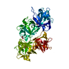
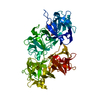
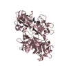
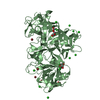
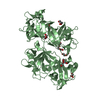
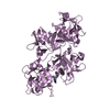
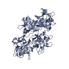
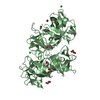
| Entry | Database: PDB / ID: 6i18 | ||||||
|---|---|---|---|---|---|---|---|
| Title | CRYSTAL STRUCTURE OF FASCIN IN COMPLEX WITH BDP-13176 | ||||||
 Components Components | Fascin | ||||||
 Keywords Keywords | STRUCTURAL PROTEIN / actin bundling / small molecule inhibition | ||||||
| Function / homology |  Function and homology information Function and homology informationmicrospike / parallel actin filament bundle assembly / regulation of microvillus assembly / microspike assembly / establishment of apical/basal cell polarity / positive regulation of extracellular matrix disassembly / cell projection membrane / positive regulation of podosome assembly / cell-cell junction assembly / podosome ...microspike / parallel actin filament bundle assembly / regulation of microvillus assembly / microspike assembly / establishment of apical/basal cell polarity / positive regulation of extracellular matrix disassembly / cell projection membrane / positive regulation of podosome assembly / cell-cell junction assembly / podosome / positive regulation of filopodium assembly / microvillus / establishment or maintenance of cell polarity / actin filament bundle assembly / positive regulation of lamellipodium assembly / ruffle / stress fiber / regulation of actin cytoskeleton organization / cell motility / filopodium / cell-cell junction / actin filament binding / cell migration / lamellipodium / actin cytoskeleton / growth cone / actin cytoskeleton organization / actin binding / Interleukin-4 and Interleukin-13 signaling / cell cortex / cytoskeleton / protein-macromolecule adaptor activity / cadherin binding / RNA binding / extracellular exosome / cytosol / cytoplasm Similarity search - Function | ||||||
| Biological species |  Homo sapiens (human) Homo sapiens (human) | ||||||
| Method |  X-RAY DIFFRACTION / SYNCHROTRON / MOLECULAR REPLACEMENT / Resolution: 1.49 Å X-RAY DIFFRACTION / SYNCHROTRON / MOLECULAR REPLACEMENT / Resolution: 1.49 Å | ||||||
 Authors Authors | Schuettelkopf, A.W. | ||||||
 Citation Citation | Journal: Bioorg.Med.Chem.Lett. / Year: 2019 Title: Structure-based design, synthesis and biological evaluation of a novel series of isoquinolone and pyrazolo[4,3-c]pyridine inhibitors of fascin 1 as potential anti-metastatic agents. Authors: Francis, S. / Croft, D. / Schuttelkopf, A.W. / Parry, C. / Pugliese, A. / Cameron, K. / Claydon, S. / Drysdale, M. / Gardner, C. / Gohlke, A. / Goodwin, G. / Gray, C.H. / Konczal, J. / ...Authors: Francis, S. / Croft, D. / Schuttelkopf, A.W. / Parry, C. / Pugliese, A. / Cameron, K. / Claydon, S. / Drysdale, M. / Gardner, C. / Gohlke, A. / Goodwin, G. / Gray, C.H. / Konczal, J. / McDonald, L. / Mezna, M. / Pannifer, A. / Paul, N.R. / Machesky, L. / McKinnon, H. / Bower, J. | ||||||
| History |
|
- Structure visualization
| Structure viewer | Molecule: MolmilJmol/JSmol |
|---|
- Downloads & links
-Download
| PDBx/mmCIF format | 6i18.cif.gz | 221.2 KB | Display | PDBx/mmCIF format |
|---|---|---|---|---|
| PDB format | pdb6i18.ent.gz | 176.4 KB | Display | PDB format |
| PDBx/mmJSON format | 6i18.json.gz | Tree view | PDBx/mmJSON format | |
| Others |  Other downloads Other downloads |
-Validation report
| Arichive directory | https://data.pdbj.org/pub/pdb/validation_reports/i1/6i18ftp://data.pdbj.org/pub/pdb/validation_reports/i1/6i18 | HTTPS FTP |
|---|
-Related structure data
| Related structure data |  6i0zC  6i10SC  6i11C  6i12C  6i13C  6i14C  6i15C  6i16C  6i17C S: Starting model for refinement C: citing same article ( |
|---|---|
| Similar structure data |

-Links
 PDBj
PDBj- Assembly
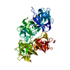
| Deposited unit | 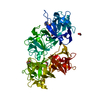
| ||||||||
|---|---|---|---|---|---|---|---|---|---|
| 1 |
| ||||||||
| Unit cell |
|
-Components
| #1: Protein | Mass: 54601.879 Da / Num. of mol.: 1 Source method: isolated from a genetically manipulated source Source: (gene. exp.) Homo sapiens (human) / Gene: FSCN1, FAN1, HSN, SNL / Plasmid: pBDDP-SPR3 / Production host:  | ||||
|---|---|---|---|---|---|
| #2: Chemical | ChemComp-ACT /   Mass: 59.044 Da / Num. of mol.: 1 / Source method: obtained synthetically / Formula: C2H3O2 Mass: 59.044 Da / Num. of mol.: 1 / Source method: obtained synthetically / Formula: C2H3O2 | ||||
| #3: Chemical | ChemComp-EDO /   Mass: 62.068 Da / Num. of mol.: 5 / Source method: obtained synthetically / Formula: C2H6O2 Mass: 62.068 Da / Num. of mol.: 5 / Source method: obtained synthetically / Formula: C2H6O2#4: Chemical | ChemComp-H0N / |   Mass: 497.376 Da / Num. of mol.: 1 / Source method: obtained synthetically / Formula: C24H22Cl2N6O2 / Feature type: SUBJECT OF INVESTIGATION Mass: 497.376 Da / Num. of mol.: 1 / Source method: obtained synthetically / Formula: C24H22Cl2N6O2 / Feature type: SUBJECT OF INVESTIGATION#5: Water | ChemComp-HOH / |  Mass: 18.015 Da / Num. of mol.: 349 / Source method: isolated from a natural source / Formula: H2O Mass: 18.015 Da / Num. of mol.: 349 / Source method: isolated from a natural source / Formula: H2O |
-Experimental details
-Experiment
| Experiment | Method: X-RAY DIFFRACTION / Number of used crystals: 1 |
|---|
- Sample preparation
| Crystal | Density Matthews: 2.36 Å3/Da / Density % sol: 47.82 % / Mosaicity: 0.214 ° |
|---|---|
| Crystal grow | Temperature: 292 K / Method: vapor diffusion, sitting drop / pH: 5 Details: 18-22% PEG 8000, 100-130 mM MgAc2, 100 mM citric acid pH 5.0 |
-Data collection
| Diffraction | Mean temperature: 100 K / Serial crystal experiment: N |
|---|---|
| Diffraction source | Source: SYNCHROTRON / Site: Diamond  / Beamline: I03 / Wavelength: 0.97625 Å / Beamline: I03 / Wavelength: 0.97625 Å |
| Detector | Type: DECTRIS PILATUS3 6M / Detector: PIXEL / Date: Jan 23, 2016 |
| Radiation | Protocol: SINGLE WAVELENGTH / Monochromatic (M) / Laue (L): M / Scattering type: x-ray |
| Radiation wavelength | Wavelength: 0.97625 Å / Relative weight: 1 |
| Reflection | Resolution: 1.49→49.35 Å / Num. obs: 84863 / % possible obs: 99.9 % / Redundancy: 6.4 % / Biso Wilson estimate: 19.368 Å2 / Rmerge(I) obs: 0.045 / Rpim(I) all: 0.021 / Rrim(I) all: 0.054 / Net I/σ(I): 18 / Num. measured all: 543840 |
| Reflection shell | Resolution: 1.49→1.53 Å / Redundancy: 6.5 % / Rmerge(I) obs: 1.014 / Mean I/σ(I) obs: 1.7 / Rpim(I) all: 0.464 / Rrim(I) all: 1.195 / % possible all: 99.9 |
- Processing
| Software |
| ||||||||||||||||||||||||||||||||||||||||||||||||||||||||||||||||||||||||||||||||||||||||||||||||||||||||||||||||||||||||||||||||||||||||||||||||||||||
|---|---|---|---|---|---|---|---|---|---|---|---|---|---|---|---|---|---|---|---|---|---|---|---|---|---|---|---|---|---|---|---|---|---|---|---|---|---|---|---|---|---|---|---|---|---|---|---|---|---|---|---|---|---|---|---|---|---|---|---|---|---|---|---|---|---|---|---|---|---|---|---|---|---|---|---|---|---|---|---|---|---|---|---|---|---|---|---|---|---|---|---|---|---|---|---|---|---|---|---|---|---|---|---|---|---|---|---|---|---|---|---|---|---|---|---|---|---|---|---|---|---|---|---|---|---|---|---|---|---|---|---|---|---|---|---|---|---|---|---|---|---|---|---|---|---|---|---|---|---|---|---|
| Refinement | Method to determine structure: MOLECULAR REPLACEMENT Starting model: 6I10 Resolution: 1.49→49.35 Å / Cor.coef. Fo:Fc: 0.971 / Cor.coef. Fo:Fc free: 0.964 / SU B: 3.176 / SU ML: 0.051 / Cross valid method: THROUGHOUT / σ(F): 0 / ESU R: 0.067 / ESU R Free: 0.068 Details: U VALUES : WITH TLS ADDED HYDROGENS HAVE BEEN ADDED IN THE RIDING POSITIONS
| ||||||||||||||||||||||||||||||||||||||||||||||||||||||||||||||||||||||||||||||||||||||||||||||||||||||||||||||||||||||||||||||||||||||||||||||||||||||
| Solvent computation | Ion probe radii: 0.8 Å / Shrinkage radii: 0.8 Å / VDW probe radii: 1.2 Å | ||||||||||||||||||||||||||||||||||||||||||||||||||||||||||||||||||||||||||||||||||||||||||||||||||||||||||||||||||||||||||||||||||||||||||||||||||||||
| Displacement parameters | Biso max: 64.29 Å2 / Biso mean: 26.229 Å2 / Biso min: 13.28 Å2
| ||||||||||||||||||||||||||||||||||||||||||||||||||||||||||||||||||||||||||||||||||||||||||||||||||||||||||||||||||||||||||||||||||||||||||||||||||||||
| Refinement step | Cycle: final / Resolution: 1.49→49.35 Å
| ||||||||||||||||||||||||||||||||||||||||||||||||||||||||||||||||||||||||||||||||||||||||||||||||||||||||||||||||||||||||||||||||||||||||||||||||||||||
| Refine LS restraints |
| ||||||||||||||||||||||||||||||||||||||||||||||||||||||||||||||||||||||||||||||||||||||||||||||||||||||||||||||||||||||||||||||||||||||||||||||||||||||
| LS refinement shell | Resolution: 1.49→1.529 Å / Total num. of bins used: 20
| ||||||||||||||||||||||||||||||||||||||||||||||||||||||||||||||||||||||||||||||||||||||||||||||||||||||||||||||||||||||||||||||||||||||||||||||||||||||
| Refinement TLS params. | Method: refined / Refine-ID: X-RAY DIFFRACTION
| ||||||||||||||||||||||||||||||||||||||||||||||||||||||||||||||||||||||||||||||||||||||||||||||||||||||||||||||||||||||||||||||||||||||||||||||||||||||
| Refinement TLS group |
|
