 Movie
Movie Controller


+ Open data

- Basic information
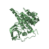
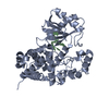
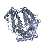
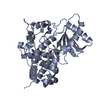
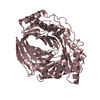
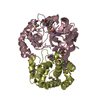
| Entry | Database: PDB / ID: 6i0s | ||||||
|---|---|---|---|---|---|---|---|
| Title | Crystal structure of DmTailor in complex with UMPNPP | ||||||
 Components Components | Terminal uridylyltransferase Tailor | ||||||
 Keywords Keywords | TRANSFERASE / Terminal uridyl transferase / nucleotidyl transferase | ||||||
| Function / homology |  Function and homology information Function and homology informationnegative regulation of pre-miRNA processing / positive regulation of miRNA catabolic process / RNA uridylyltransferase / RNA uridylyltransferase activity / RNA 3'-end processing / regulatory ncRNA-mediated gene silencing / ATP binding / metal ion binding / cytoplasm Similarity search - Function | ||||||
| Biological species |  | ||||||
| Method |  X-RAY DIFFRACTION / SYNCHROTRON / MOLECULAR REPLACEMENT / Resolution: 1.9 Å X-RAY DIFFRACTION / SYNCHROTRON / MOLECULAR REPLACEMENT / Resolution: 1.9 Å | ||||||
 Authors Authors | Kroupova, A. / Ivascu, A. / Jinek, M. | ||||||
 Citation Citation | Journal: Nucleic Acids Res. / Year: 2019 Title: Structural basis for acceptor RNA substrate selectivity of the 3' terminal uridylyl transferase Tailor. Authors: Kroupova, A. / Ivascu, A. / Reimao-Pinto, M.M. / Ameres, S.L. / Jinek, M. | ||||||
| History |
|
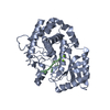
- Structure visualization
| Structure viewer | Molecule: MolmilJmol/JSmol |
|---|
- Downloads & links
-Download
| PDBx/mmCIF format | 6i0s.cif.gz | 220.8 KB | Display | PDBx/mmCIF format |
|---|---|---|---|---|
| PDB format | pdb6i0s.ent.gz | 178.4 KB | Display | PDB format |
| PDBx/mmJSON format | 6i0s.json.gz | Tree view | PDBx/mmJSON format | |
| Others |  Other downloads Other downloads |
-Validation report
| Arichive directory | https://data.pdbj.org/pub/pdb/validation_reports/i0/6i0sftp://data.pdbj.org/pub/pdb/validation_reports/i0/6i0s | HTTPS FTP |
|---|
-Related structure data
| Related structure data |  6i0tC  6i0uC 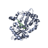 6i0vC  5w0bS C: citing same article ( S: Starting model for refinement |
|---|---|
| Similar structure data |


-Links
 PDBj
PDBj- Assembly
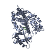
| Deposited unit | 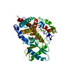
| ||||||||
|---|---|---|---|---|---|---|---|---|---|
| 1 |
| ||||||||
| Unit cell |
|
-Components
| #1: Protein | Mass: 44259.301 Da / Num. of mol.: 1 Source method: isolated from a genetically manipulated source Source: (gene. exp.)  Spodoptera frugiperda (fall armyworm) / References: UniProt: Q9VI58, RNA uridylyltransferase Spodoptera frugiperda (fall armyworm) / References: UniProt: Q9VI58, RNA uridylyltransferase |
|---|---|
| #2: Chemical | ChemComp-2KH / 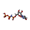  Mass: 483.156 Da / Num. of mol.: 1 / Source method: obtained synthetically / Formula: C9H16N3O14P3 Mass: 483.156 Da / Num. of mol.: 1 / Source method: obtained synthetically / Formula: C9H16N3O14P3 |
| #3: Chemical | ChemComp-MG /   Mass: 24.305 Da / Num. of mol.: 1 / Source method: isolated from a natural source / Formula: Mg Mass: 24.305 Da / Num. of mol.: 1 / Source method: isolated from a natural source / Formula: Mg |
| #4: Water | ChemComp-HOH /  Mass: 18.015 Da / Num. of mol.: 221 / Source method: isolated from a natural source / Formula: H2O Mass: 18.015 Da / Num. of mol.: 221 / Source method: isolated from a natural source / Formula: H2O |
-Experimental details
-Experiment
| Experiment | Method: X-RAY DIFFRACTION / Number of used crystals: 1 |
|---|
- Sample preparation
| Crystal | Density Matthews: 2.27 Å3/Da / Density % sol: 45.91 % |
|---|---|
| Crystal grow | Temperature: 293 K / Method: vapor diffusion, hanging drop / pH: 7 / Details: 20 % (v/v) glycerol ethoxylate |
-Data collection
| Diffraction | Mean temperature: 100 K / Serial crystal experiment: N |
|---|---|
| Diffraction source | Source: SYNCHROTRON / Site: SLS  / Beamline: X06DA / Wavelength: 1.00767 Å / Beamline: X06DA / Wavelength: 1.00767 Å |
| Detector | Type: DECTRIS PILATUS 2M-F / Detector: PIXEL / Date: Nov 3, 2017 |
| Radiation | Protocol: SINGLE WAVELENGTH / Monochromatic (M) / Laue (L): M / Scattering type: x-ray |
| Radiation wavelength | Wavelength: 1.00767 Å / Relative weight: 1 |
| Reflection | Resolution: 1.9→44.77 Å / Num. obs: 29527 / % possible obs: 100 % / Redundancy: 19.6 % / CC1/2: 0.999 / Rsym value: 0.119 / Net I/σ(I): 15.81 |
| Reflection shell | Resolution: 1.9→2.01 Å / Redundancy: 19 % / Mean I/σ(I) obs: 1.4 / CC1/2: 0.534 / Rsym value: 1.55 / % possible all: 100 |
- Processing
| Software |
| ||||||||||||||||||||||||||||||||||||||||||||||||||||||||||||||||||||||||||||||||||||
|---|---|---|---|---|---|---|---|---|---|---|---|---|---|---|---|---|---|---|---|---|---|---|---|---|---|---|---|---|---|---|---|---|---|---|---|---|---|---|---|---|---|---|---|---|---|---|---|---|---|---|---|---|---|---|---|---|---|---|---|---|---|---|---|---|---|---|---|---|---|---|---|---|---|---|---|---|---|---|---|---|---|---|---|---|---|
| Refinement | Method to determine structure: MOLECULAR REPLACEMENT Starting model: 5W0B Resolution: 1.9→44.768 Å / SU ML: 0.2 / Cross valid method: FREE R-VALUE / σ(F): 1.35 / Phase error: 19.9
| ||||||||||||||||||||||||||||||||||||||||||||||||||||||||||||||||||||||||||||||||||||
| Solvent computation | Shrinkage radii: 0.9 Å / VDW probe radii: 1.11 Å | ||||||||||||||||||||||||||||||||||||||||||||||||||||||||||||||||||||||||||||||||||||
| Refinement step | Cycle: LAST / Resolution: 1.9→44.768 Å
| ||||||||||||||||||||||||||||||||||||||||||||||||||||||||||||||||||||||||||||||||||||
| Refine LS restraints |
| ||||||||||||||||||||||||||||||||||||||||||||||||||||||||||||||||||||||||||||||||||||
| LS refinement shell |
|
