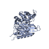
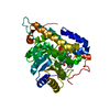
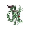
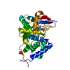
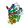
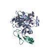
Entry Database : PDB / ID : 6hziTitle Apo structure of TP domain from Burkholderia pseudomallei penicillin-binding protein 3 Peptidoglycan D,D-transpeptidase FtsI Keywords / / / Function / homology Function Domain/homology Component
/ / / / / / / / / / / / / / / / / / / / / / / / / / / / / / / / / / / / / / / / Biological species Burkholderia pseudomallei (bacteria)Method / / / Resolution : 2.29 Å Authors Bellini, D. / Koekemoer, L. / Newman, H. / Dowson, C.G. Funding support Organization Grant number Country Medical Research Council (United Kingdom) grant.MR/P007503/1
Journal : To Be Published Title : Apo structure of TP domain from Burkholderia pseudomallei penicillin-binding protein 3Authors : Bellini, D. / Koekemoer, L. / Newman, H. / Dowson, C.G. History Deposition Oct 23, 2018 Deposition site / Processing site Revision 1.0 Nov 20, 2019 Provider / Type Revision 1.1 Jan 24, 2024 Group / Database references / Refinement descriptionCategory chem_comp_atom / chem_comp_bond ... chem_comp_atom / chem_comp_bond / database_2 / pdbx_initial_refinement_model Item / _database_2.pdbx_database_accession
Show all Show less
 Movie
Movie Controller

 Yorodumi
 Open data
 Basic information
 Components
 Keywords
 Function and homology information
 Burkholderia pseudomallei (bacteria)
 X-RAY DIFFRACTION /
  Authors
 United Kingdom, 1items
  Citation
 Structure visualization
 Downloads & links
 Other downloads
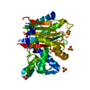


 PDBj
PDBj
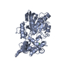
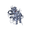
 Assembly
 Mass: 18.015 Da / Num. of mol.: 56 / Source method: isolated from a natural source / Formula: H2O
 Sample preparation
 Processing