 Movie
Movie Controller

[English] 日本語
 Yorodumi
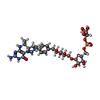
- PDB-6hux: HmdII from Methanocaldococcus jannaschii reconstitued with Fe-gua... -
+ Open data

- Basic information
| Entry | Database: PDB / ID: 6hux | |||||||||
|---|---|---|---|---|---|---|---|---|---|---|
| Title | HmdII from Methanocaldococcus jannaschii reconstitued with Fe-guanylylpyridinol (FeGP) cofactor and co-crystallized with methenyl-tetrahydromethanopterin at 2.5 A resolution | |||||||||
 Components Components | H(2)-forming methylenetetrahydromethanopterin dehydrogenase-related protein MJ1338 | |||||||||
 Keywords Keywords | OXIDOREDUCTASE / Hydrogenase / H2-activation / Lateral gene-transfer / cofactor biosynthesis / tetrahydromethanopterin / paralog / methanogen / metalloenzyme | |||||||||
| Function / homology |  Function and homology information Function and homology information | |||||||||
| Biological species |   Methanocaldococcus jannaschii DSM 2661 (archaea) Methanocaldococcus jannaschii DSM 2661 (archaea) | |||||||||
| Method |  X-RAY DIFFRACTION / SYNCHROTRON / MOLECULAR REPLACEMENT / Resolution: 2.5 Å X-RAY DIFFRACTION / SYNCHROTRON / MOLECULAR REPLACEMENT / Resolution: 2.5 Å | |||||||||
 Authors Authors | Watanabe, T. / Wagner, T. / Huang, G. / Kahnt, J. / Ataka, K. / Ermler, U. / Shima, S. | |||||||||
| Funding support |  Germany, Germany,  China, 2items China, 2items
| |||||||||
 Citation Citation | Journal: Angew. Chem. Int. Ed. Engl. / Year: 2019 Title: The Bacterial [Fe]-Hydrogenase Paralog HmdII Uses Tetrahydrofolate Derivatives as Substrates. Authors: Watanabe, T. / Wagner, T. / Huang, G. / Kahnt, J. / Ataka, K. / Ermler, U. / Shima, S. | |||||||||
| History |
|
- Structure visualization
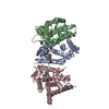
| Structure viewer | Molecule: MolmilJmol/JSmol |
|---|
- Downloads & links
-Download
| PDBx/mmCIF format | 6hux.cif.gz | 165.5 KB | Display | PDBx/mmCIF format |
|---|---|---|---|---|
| PDB format | pdb6hux.ent.gz | 130.1 KB | Display | PDB format |
| PDBx/mmJSON format | 6hux.json.gz | Tree view | PDBx/mmJSON format | |
| Others |  Other downloads Other downloads |
-Validation report
| Arichive directory | https://data.pdbj.org/pub/pdb/validation_reports/hu/6huxftp://data.pdbj.org/pub/pdb/validation_reports/hu/6hux | HTTPS FTP |
|---|
-Related structure data
| Related structure data |  6huyC  6huzC  4yt4S S: Starting model for refinement C: citing same article ( |
|---|---|
| Similar structure data |


-Links
 PDBj
PDBj


- Assembly
| Deposited unit | 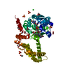
| |||||||||||||||
|---|---|---|---|---|---|---|---|---|---|---|---|---|---|---|---|---|
| 1 | 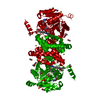
| |||||||||||||||
| Unit cell |
| |||||||||||||||
| Components on special symmetry positions |
|
-Components
-Protein , 1 types, 1 molecules A
| #1: Protein | Mass: 40663.848 Da / Num. of mol.: 1 Source method: isolated from a genetically manipulated source Details: / Source: (gene. exp.) Methanocaldococcus jannaschii DSM 2661 (archaea)Tissue: / / Cell: / / Cell line: / / Gene: MJ1338 / Organ: / / Details (production host): / / Cell (production host): / / Organ (production host): / / Production host:  References: UniProt: Q58734, 5,10-methenyltetrahydromethanopterin hydrogenase |
|---|
-Non-polymers , 9 types, 77 molecules 
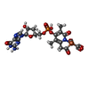


| #2: Chemical | ChemComp-ACT /  Mass: 59.044 Da / Num. of mol.: 9 / Source method: obtained synthetically / Formula: C2H3O2 Mass: 59.044 Da / Num. of mol.: 9 / Source method: obtained synthetically / Formula: C2H3O2#3: Chemical | ChemComp-FE9 / |  Mass: 686.323 Da / Num. of mol.: 1 / Source method: obtained synthetically / Formula: C21H23FeN6O13PS Mass: 686.323 Da / Num. of mol.: 1 / Source method: obtained synthetically / Formula: C21H23FeN6O13PS#4: Chemical | ChemComp-NA / |  Mass: 22.990 Da / Num. of mol.: 1 / Source method: obtained synthetically / Formula: Na Mass: 22.990 Da / Num. of mol.: 1 / Source method: obtained synthetically / Formula: Na#5: Chemical | ChemComp-E4M / |  Mass: 787.685 Da / Num. of mol.: 1 / Source method: obtained synthetically / Formula: C31H44N6O16P Mass: 787.685 Da / Num. of mol.: 1 / Source method: obtained synthetically / Formula: C31H44N6O16P#6: Chemical | ChemComp-MG / |  Mass: 24.305 Da / Num. of mol.: 1 / Source method: obtained synthetically / Formula: Mg Mass: 24.305 Da / Num. of mol.: 1 / Source method: obtained synthetically / Formula: Mg#7: Chemical | ChemComp-SO4 /  Mass: 96.063 Da / Num. of mol.: 7 / Source method: obtained synthetically / Formula: SO4 Mass: 96.063 Da / Num. of mol.: 7 / Source method: obtained synthetically / Formula: SO4#8: Chemical |  Mass: 62.068 Da / Num. of mol.: 3 / Source method: obtained synthetically / Formula: C2H6O2 Mass: 62.068 Da / Num. of mol.: 3 / Source method: obtained synthetically / Formula: C2H6O2#9: Chemical | ChemComp-CL / |  Mass: 35.453 Da / Num. of mol.: 1 / Source method: obtained synthetically / Formula: Cl Mass: 35.453 Da / Num. of mol.: 1 / Source method: obtained synthetically / Formula: Cl#10: Water | ChemComp-HOH / | Mass: 18.015 Da / Num. of mol.: 53 / Source method: isolated from a natural source / Formula: H2O |
|---|
-Experimental details
-Experiment
| Experiment | Method: X-RAY DIFFRACTION / Number of used crystals: 1 |
|---|
- Sample preparation
| Crystal | Density Matthews: 2.75 Å3/Da / Density % sol: 55.25 % / Description: transparent flower shape |
|---|---|
| Crystal grow | Temperature: 281 K / Method: vapor diffusion, sitting drop / pH: 5.5 Details: HmdII from Methanocaldococcus jannaschii reconstituted with Fe-guanylylpyridinol cofactor was cocrystallized with methenyl-tetrahydromethanopterin using the sitting drop vapor diffusion ...Details: HmdII from Methanocaldococcus jannaschii reconstituted with Fe-guanylylpyridinol cofactor was cocrystallized with methenyl-tetrahydromethanopterin using the sitting drop vapor diffusion method under N2/H2 (95%/5%) in red light condition. 20 mg/ml of reconstituted enzyme in 25 mM Tris pH 7.5, 5% glycerol, 150 mM NaCl, 2 mM DTT and 3 mM methenyl-tetrahydromethanopterin was spotted on a 96-well 2-drop MRC Crystallization Plates (Molecular Dimensions, Suffolk, UK) with a ratio of 0.7 ul of protein and 0.7 ul of reservoir solution. After several weeks, crystals appeared in 2 M LiSO4, 100 mM Sodium acetate pH 5.5, 100 mM MgSO4 and 5% v/v PEG 400. jHmdII crystal was cryoprotected in its mother liquor supplemented with 30% ethylene glycol before freezing in liquid nitrogen. Temp details: The temperature fluctuation was +/- 1 degree |
-Data collection
| Diffraction | Mean temperature: 100 K / Serial crystal experiment: N |
|---|---|
| Diffraction source | Source: SYNCHROTRON / Site: ESRF  / Beamline: BM30A / Wavelength: 0.9799 Å / Beamline: BM30A / Wavelength: 0.9799 Å |
| Detector | Type: ADSC QUANTUM 315r / Detector: CCD / Date: Apr 22, 2017 |
| Radiation | Protocol: SINGLE WAVELENGTH / Monochromatic (M) / Laue (L): M / Scattering type: x-ray |
| Radiation wavelength | Wavelength: 0.9799 Å / Relative weight: 1 |
| Reflection | Resolution: 2.5→43.73 Å / Num. obs: 15610 / % possible obs: 100 % / Redundancy: 9.8 % / Biso Wilson estimate: 64.85 Å2 / CC1/2: 0.997 / Rmerge(I) obs: 0.122 / Rpim(I) all: 0.041 / Rrim(I) all: 0.129 / Net I/σ(I): 13.1 |
| Reflection shell | Resolution: 2.5→2.64 Å / Redundancy: 9.9 % / Rmerge(I) obs: 1.341 / Mean I/σ(I) obs: 2.2 / Num. unique obs: 2224 / CC1/2: 0.686 / Rpim(I) all: 0.447 / Rrim(I) all: 1.415 / % possible all: 99.9 |
- Processing
| Software |
| |||||||||||||||||||||||||||||||||||||||||||||||||||||||||||||||||||||||||||||||||||||||||||||||||||||||||||||||||||||||||||||||||||||||||||||||||||||||||||||||||||||||||||||||
|---|---|---|---|---|---|---|---|---|---|---|---|---|---|---|---|---|---|---|---|---|---|---|---|---|---|---|---|---|---|---|---|---|---|---|---|---|---|---|---|---|---|---|---|---|---|---|---|---|---|---|---|---|---|---|---|---|---|---|---|---|---|---|---|---|---|---|---|---|---|---|---|---|---|---|---|---|---|---|---|---|---|---|---|---|---|---|---|---|---|---|---|---|---|---|---|---|---|---|---|---|---|---|---|---|---|---|---|---|---|---|---|---|---|---|---|---|---|---|---|---|---|---|---|---|---|---|---|---|---|---|---|---|---|---|---|---|---|---|---|---|---|---|---|---|---|---|---|---|---|---|---|---|---|---|---|---|---|---|---|---|---|---|---|---|---|---|---|---|---|---|---|---|---|---|---|---|
| Refinement | Method to determine structure: MOLECULAR REPLACEMENT Starting model: 4YT4 Resolution: 2.5→38.97 Å / Cor.coef. Fo:Fc: 0.959 / Cor.coef. Fo:Fc free: 0.912 / Cross valid method: THROUGHOUT / σ(F): 0 / SU R Blow DPI: 0.372 / SU Rfree Blow DPI: 0.236 Details: The last refinement cycle was performed with hydrogens in riding position. The hydrogens have been removed from the deposited model.
| |||||||||||||||||||||||||||||||||||||||||||||||||||||||||||||||||||||||||||||||||||||||||||||||||||||||||||||||||||||||||||||||||||||||||||||||||||||||||||||||||||||||||||||||
| Displacement parameters | Biso mean: 64.84 Å2
| |||||||||||||||||||||||||||||||||||||||||||||||||||||||||||||||||||||||||||||||||||||||||||||||||||||||||||||||||||||||||||||||||||||||||||||||||||||||||||||||||||||||||||||||
| Refine analyze | Luzzati coordinate error obs: 0.27 Å | |||||||||||||||||||||||||||||||||||||||||||||||||||||||||||||||||||||||||||||||||||||||||||||||||||||||||||||||||||||||||||||||||||||||||||||||||||||||||||||||||||||||||||||||
| Refinement step | Cycle: 1 / Resolution: 2.5→38.97 Å
| |||||||||||||||||||||||||||||||||||||||||||||||||||||||||||||||||||||||||||||||||||||||||||||||||||||||||||||||||||||||||||||||||||||||||||||||||||||||||||||||||||||||||||||||
| Refine LS restraints |
| |||||||||||||||||||||||||||||||||||||||||||||||||||||||||||||||||||||||||||||||||||||||||||||||||||||||||||||||||||||||||||||||||||||||||||||||||||||||||||||||||||||||||||||||
| LS refinement shell | Resolution: 2.5→2.67 Å / Total num. of bins used: 8
| |||||||||||||||||||||||||||||||||||||||||||||||||||||||||||||||||||||||||||||||||||||||||||||||||||||||||||||||||||||||||||||||||||||||||||||||||||||||||||||||||||||||||||||||
| Refinement TLS params. | Method: refined / Refine-ID: X-RAY DIFFRACTION
| |||||||||||||||||||||||||||||||||||||||||||||||||||||||||||||||||||||||||||||||||||||||||||||||||||||||||||||||||||||||||||||||||||||||||||||||||||||||||||||||||||||||||||||||
| Refinement TLS group |
|
