 Movie
Movie Controller

[English] 日本語
 Yorodumi
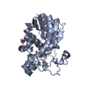
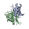
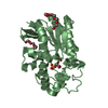
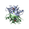
- PDB-6hsy: Two-phospholipid-bound crystal structure of the substrate-binding... -
+ Open data

- Basic information
| Entry | Database: PDB / ID: 6hsy | |||||||||
|---|---|---|---|---|---|---|---|---|---|---|
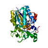
| Title | Two-phospholipid-bound crystal structure of the substrate-binding protein Ttg2D from Pseudomonas aeruginosa | |||||||||
 Components Components | Toluene tolerance protein Ttg2D | |||||||||
 Keywords Keywords | LIPID TRANSPORT / substrate-binding protein / two diacyl lipids / Ttg2D / MlaC | |||||||||
| Function / homology |  Function and homology information Function and homology informationTgt2/MlaC / Tgt2/MlaC superfamily / Toluene tolerance Ttg2/phospholipid-binding protein MlaC / MlaC protein / Nuclear Transport Factor 2; Chain: A, / Roll / Alpha Beta Similarity search - Domain/homology | |||||||||
| Biological species |  Pseudomonas aeruginosa PAO1 (bacteria) Pseudomonas aeruginosa PAO1 (bacteria) | |||||||||
| Method |  X-RAY DIFFRACTION / SYNCHROTRON / MOLECULAR REPLACEMENT / Resolution: 2.53 Å X-RAY DIFFRACTION / SYNCHROTRON / MOLECULAR REPLACEMENT / Resolution: 2.53 Å | |||||||||
 Authors Authors | Costenaro, L. / Conchillo-Sole, O. / Daura, X. | |||||||||
| Funding support |  Spain, 2items Spain, 2items
| |||||||||
 Citation Citation | Journal: Commun Biol / Year: 2021 Title: The Pseudomonas aeruginosa substrate-binding protein Ttg2D functions as a general glycerophospholipid transporter across the periplasm. Authors: Yero, D. / Diaz-Lobo, M. / Costenaro, L. / Conchillo-Sole, O. / Mayo, A. / Ferrer-Navarro, M. / Vilaseca, M. / Gibert, I. / Daura, X. | |||||||||
| History |
|
- Structure visualization
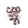
| Structure viewer | Molecule: MolmilJmol/JSmol |
|---|
- Downloads & links
-Download
| PDBx/mmCIF format | 6hsy.cif.gz | 95.8 KB | Display | PDBx/mmCIF format |
|---|---|---|---|---|
| PDB format | pdb6hsy.ent.gz | 72.4 KB | Display | PDB format |
| PDBx/mmJSON format | 6hsy.json.gz | Tree view | PDBx/mmJSON format | |
| Others |  Other downloads Other downloads |
-Validation report
| Arichive directory | https://data.pdbj.org/pub/pdb/validation_reports/hs/6hsyftp://data.pdbj.org/pub/pdb/validation_reports/hs/6hsy | HTTPS FTP |
|---|
-Related structure data
| Related structure data |  2qguS S: Starting model for refinement |
|---|---|
| Similar structure data |


-Links
 PDBj
PDBj- Assembly
| Deposited unit | 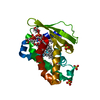
| ||||||||||||
|---|---|---|---|---|---|---|---|---|---|---|---|---|---|
| 1 |
| ||||||||||||
| Unit cell |
|
-Components
-Protein , 1 types, 1 molecules A
| #1: Protein | Mass: 23016.129 Da / Num. of mol.: 1 Source method: isolated from a genetically manipulated source Details: Expression tags:N-term: MC-term: KLAAALEHHHHHH / Source: (gene. exp.) Pseudomonas aeruginosa PAO1 (bacteria) / Gene: PA4453 / Plasmid: pET28b / Production host: |
|---|
-Non-polymers , 5 types, 28 molecules 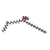
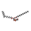


| #2: Chemical | ChemComp-GOT /  Mass: 734.981 Da / Num. of mol.: 1 / Source method: obtained synthetically / Formula: C39H75O10P Mass: 734.981 Da / Num. of mol.: 1 / Source method: obtained synthetically / Formula: C39H75O10P | ||||
|---|---|---|---|---|---|
| #3: Chemical | ChemComp-H3T /  Mass: 734.981 Da / Num. of mol.: 1 / Source method: obtained synthetically / Formula: C39H75O10P Mass: 734.981 Da / Num. of mol.: 1 / Source method: obtained synthetically / Formula: C39H75O10P | ||||
| #4: Chemical |  Mass: 92.094 Da / Num. of mol.: 2 / Source method: obtained synthetically / Formula: C3H8O3 Mass: 92.094 Da / Num. of mol.: 2 / Source method: obtained synthetically / Formula: C3H8O3#5: Chemical |  Mass: 96.063 Da / Num. of mol.: 3 / Source method: obtained synthetically / Formula: SO4 Mass: 96.063 Da / Num. of mol.: 3 / Source method: obtained synthetically / Formula: SO4#6: Water | ChemComp-HOH / | Mass: 18.015 Da / Num. of mol.: 21 / Source method: isolated from a natural source / Formula: H2O |
-Experimental details
-Experiment
| Experiment | Method: X-RAY DIFFRACTION / Number of used crystals: 1 |
|---|
- Sample preparation
| Crystal | Density Matthews: 3.71 Å3/Da / Density % sol: 66.83 % |
|---|---|
| Crystal grow | Temperature: 293.15 K / Method: vapor diffusion, hanging drop Details: 0.17 M Ammonium sulfate, 25.5% w/v PEG 4000, 15% v/v Glycerol |
-Data collection
| Diffraction | Mean temperature: 100 K / Serial crystal experiment: N |
|---|---|
| Diffraction source | Source: SYNCHROTRON / Site: ESRF  / Beamline: ID23-1 / Wavelength: 0.980227 Å / Beamline: ID23-1 / Wavelength: 0.980227 Å |
| Detector | Type: DECTRIS PILATUS 6M-F / Detector: PIXEL / Date: Jun 28, 2013 |
| Radiation | Monochromator: Silicon (1 1 1) channel-cut / Protocol: SINGLE WAVELENGTH / Monochromatic (M) / Laue (L): M / Scattering type: x-ray |
| Radiation wavelength | Wavelength: 0.980227 Å / Relative weight: 1 |
| Reflection | Resolution: 2.53→62.32 Å / Num. obs: 11489 / % possible obs: 99.6 % / Redundancy: 6.2 % / Biso Wilson estimate: 51.33 Å2 / CC1/2: 0.996 / Rmerge(I) obs: 0.124 / Rpim(I) all: 0.053 / Rrim(I) all: 0.135 / Net I/σ(I): 8.2 |
| Reflection shell | Resolution: 2.53→2.64 Å / Redundancy: 4.6 % / Rmerge(I) obs: 0.707 / Mean I/σ(I) obs: 2 / Num. unique obs: 1349 / CC1/2: 0.717 / Rpim(I) all: 0.352 / Rrim(I) all: 0.795 / % possible all: 98 |
- Processing
| Software |
| |||||||||||||||||||||||||||||||||||
|---|---|---|---|---|---|---|---|---|---|---|---|---|---|---|---|---|---|---|---|---|---|---|---|---|---|---|---|---|---|---|---|---|---|---|---|---|
| Refinement | Method to determine structure: MOLECULAR REPLACEMENT Starting model: Poly-A homology model based on 2QGU Resolution: 2.53→62.32 Å / SU ML: 0.3451 / Cross valid method: THROUGHOUT / σ(F): 1.34 / Phase error: 29.8051 / Stereochemistry target values: ML
| |||||||||||||||||||||||||||||||||||
| Solvent computation | Shrinkage radii: 0.9 Å / VDW probe radii: 1.11 Å / Solvent model: FLAT BULK SOLVENT MODEL | |||||||||||||||||||||||||||||||||||
| Displacement parameters | Biso mean: 68.04 Å2 | |||||||||||||||||||||||||||||||||||
| Refinement step | Cycle: LAST / Resolution: 2.53→62.32 Å
| |||||||||||||||||||||||||||||||||||
| Refine LS restraints |
| |||||||||||||||||||||||||||||||||||
| LS refinement shell |
|
