 Movie
Movie Controller


+ Open data

- Basic information
| Entry | Database: PDB / ID: 6hsw | ||||||||||||
|---|---|---|---|---|---|---|---|---|---|---|---|---|---|
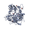
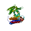
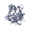
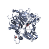
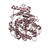
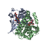
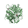
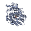
| Title | A CE15 glucuronoyl esterase from Teredinibacter turnerae T7901 | ||||||||||||
 Components Components | Carbohydrate esterase family 15 domain protein | ||||||||||||
 Keywords Keywords | HYDROLASE / CE15 / carbohydrate esterase / glucuronoyl esterase / xylan | ||||||||||||
| Function / homology | Glucuronyl esterase, fungi / Alpha/Beta hydrolase fold, catalytic domain / Alpha/Beta hydrolase fold / Rossmann fold / 3-Layer(aba) Sandwich / Alpha Beta / BROMIDE ION / DI(HYDROXYETHYL)ETHER / Carbohydrate esterase family 15 domain protein Function and homology information Function and homology information | ||||||||||||
| Biological species |  Teredinibacter turnerae T7901 (bacteria) Teredinibacter turnerae T7901 (bacteria) | ||||||||||||
| Method |  X-RAY DIFFRACTION / SYNCHROTRON / SAD / Resolution: 2.14734381231 Å X-RAY DIFFRACTION / SYNCHROTRON / SAD / Resolution: 2.14734381231 Å | ||||||||||||
 Authors Authors | Mazurkewich, S. / Lo Leggio, L. / Navarro Poulsen, J.C. / Larsbrink, J. | ||||||||||||
| Funding support |  Sweden, Sweden,  Denmark, 3items Denmark, 3items
| ||||||||||||
 Citation Citation | Journal: J.Biol.Chem. / Year: 2019 Title: Structure-function analyses reveal that a glucuronoyl esterase fromTeredinibacter turneraeinteracts with carbohydrates and aromatic compounds. Authors: Arnling Baath, J. / Mazurkewich, S. / Poulsen, J.N. / Olsson, L. / Lo Leggio, L. / Larsbrink, J. | ||||||||||||
| History |
|
- Structure visualization
| Structure viewer | Molecule: MolmilJmol/JSmol |
|---|
- Downloads & links
-Download
| PDBx/mmCIF format | 6hsw.cif.gz | 596.2 KB | Display | PDBx/mmCIF format |
|---|---|---|---|---|
| PDB format | pdb6hsw.ent.gz | 402.2 KB | Display | PDB format |
| PDBx/mmJSON format | 6hsw.json.gz | Tree view | PDBx/mmJSON format | |
| Others |  Other downloads Other downloads |
-Validation report
| Arichive directory | https://data.pdbj.org/pub/pdb/validation_reports/hs/6hswftp://data.pdbj.org/pub/pdb/validation_reports/hs/6hsw | HTTPS FTP |
|---|
-Related structure data
| Similar structure data |
|---|


-Links
 PDBj
PDBj

- Assembly
| Deposited unit | 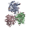
| ||||||||||||
|---|---|---|---|---|---|---|---|---|---|---|---|---|---|
| 1 | 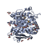
| ||||||||||||
| 2 | 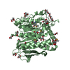
| ||||||||||||
| 3 | 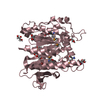
| ||||||||||||
| Unit cell |
| ||||||||||||
| Components on special symmetry positions |
|
-Components
-Protein , 1 types, 3 molecules ABC
| #1: Protein | Mass: 48081.297 Da / Num. of mol.: 3 Source method: isolated from a genetically manipulated source Source: (gene. exp.) Teredinibacter turnerae T7901 (bacteria)Gene: TERTU_0517 / Plasmid: Modified pET28-a / Details (production host): contains TEV protease site / Production host: |
|---|
-Non-polymers , 6 types, 859 molecules 


| #2: Chemical |  Mass: 238.278 Da / Num. of mol.: 2 / Source method: obtained synthetically / Formula: C10H22O6 / Comment: precipitant*YM Mass: 238.278 Da / Num. of mol.: 2 / Source method: obtained synthetically / Formula: C10H22O6 / Comment: precipitant*YM#3: Chemical | ChemComp-EDO /  Mass: 62.068 Da / Num. of mol.: 20 / Source method: obtained synthetically / Formula: C2H6O2 Mass: 62.068 Da / Num. of mol.: 20 / Source method: obtained synthetically / Formula: C2H6O2#4: Chemical | ChemComp-GOL /  Mass: 92.094 Da / Num. of mol.: 13 / Source method: obtained synthetically / Formula: C3H8O3 Mass: 92.094 Da / Num. of mol.: 13 / Source method: obtained synthetically / Formula: C3H8O3#5: Chemical |  Mass: 79.904 Da / Num. of mol.: 2 / Source method: obtained synthetically / Formula: Br Mass: 79.904 Da / Num. of mol.: 2 / Source method: obtained synthetically / Formula: Br#6: Chemical | ChemComp-PEG / |  Mass: 106.120 Da / Num. of mol.: 1 / Source method: obtained synthetically / Formula: C4H10O3 Mass: 106.120 Da / Num. of mol.: 1 / Source method: obtained synthetically / Formula: C4H10O3#7: Water | ChemComp-HOH / | Mass: 18.015 Da / Num. of mol.: 821 / Source method: isolated from a natural source / Formula: H2O |
|---|
-Details
| Has protein modification | Y |
|---|
-Experimental details
-Experiment
| Experiment | Method: X-RAY DIFFRACTION / Number of used crystals: 1 |
|---|
- Sample preparation
| Crystal | Density Matthews: 3.06 Å3/Da / Density % sol: 59.82 % |
|---|---|
| Crystal grow | Temperature: 298 K / Method: vapor diffusion, sitting drop / pH: 6.5 Details: Enzyme mixed 50/50 with reservoir solution containing Morpheus screen solution with 0.09 M halogens (0.3M Sodium fluoride; 0.3M Sodium bromide; 0.3M Sodium iodide), 0.1 M Buffer system 1 ...Details: Enzyme mixed 50/50 with reservoir solution containing Morpheus screen solution with 0.09 M halogens (0.3M Sodium fluoride; 0.3M Sodium bromide; 0.3M Sodium iodide), 0.1 M Buffer system 1 (Imidazole; MES) , and 50% v/v Precipitant mix 2 (40% v/v Ethylene glycol; 20 % w/v PEG8000) Temp details: Room Temperature |
-Data collection
| Diffraction | Mean temperature: 300 K / Serial crystal experiment: N |
|---|---|
| Diffraction source | Source: SYNCHROTRON / Site: PETRA III, DESY  / Beamline: P11 / Wavelength: 0.9795 Å / Beamline: P11 / Wavelength: 0.9795 Å |
| Detector | Type: DECTRIS PILATUS 6M-F / Detector: PIXEL / Date: Aug 26, 2017 |
| Radiation | Protocol: SINGLE WAVELENGTH / Monochromatic (M) / Laue (L): M / Scattering type: x-ray |
| Radiation wavelength | Wavelength: 0.9795 Å / Relative weight: 1 |
| Reflection | Resolution: 2.147→44.81 Å / Num. obs: 92246 / % possible obs: 99.79 % / Redundancy: 20.3 % / Biso Wilson estimate: 39.8514198624 Å2 / CC1/2: 0.995 / Rmerge(I) obs: 0.1978 / Rpim(I) all: 0.04459 / Rrim(I) all: 0.2028 / Net I/σ(I): 13.86 |
| Reflection shell | Resolution: 2.147→2.224 Å / Redundancy: 19.6 % / Mean I/σ(I) obs: 1.37 / Num. unique obs: 9051 / CC1/2: 0.634 / Rpim(I) all: 0.4095 / % possible all: 98.96 |
- Processing
| Software |
| ||||||||||||||||||||||||||||||||||||||||||||||||||||||||
|---|---|---|---|---|---|---|---|---|---|---|---|---|---|---|---|---|---|---|---|---|---|---|---|---|---|---|---|---|---|---|---|---|---|---|---|---|---|---|---|---|---|---|---|---|---|---|---|---|---|---|---|---|---|---|---|---|---|
| Refinement | Method to determine structure: SAD / Resolution: 2.14734381231→44.81 Å / SU ML: 0.254587693418 / Cross valid method: FREE R-VALUE / σ(F): 1.34354230713 / Phase error: 23.1248739467
| ||||||||||||||||||||||||||||||||||||||||||||||||||||||||
| Solvent computation | Shrinkage radii: 0.9 Å / VDW probe radii: 1.11 Å | ||||||||||||||||||||||||||||||||||||||||||||||||||||||||
| Displacement parameters | Biso mean: 50.6282588945 Å2 | ||||||||||||||||||||||||||||||||||||||||||||||||||||||||
| Refinement step | Cycle: LAST / Resolution: 2.14734381231→44.81 Å
| ||||||||||||||||||||||||||||||||||||||||||||||||||||||||
| Refine LS restraints |
| ||||||||||||||||||||||||||||||||||||||||||||||||||||||||
| LS refinement shell |
|
