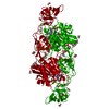
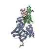
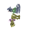
Entry Database : PDB / ID : 6hsnTitle Crystal structure of the ternary complex of GephE-ADP-GABA(A) receptor derived peptide Gamma-aminobutyric acid receptor subunit alpha-3 Gephyrin Keywords / / / / Function / homology Function Domain/homology Component
/ / / / / / / / / / / / / / / / / / / / / / / / / / / / / / / / / / / / / / / / / / / / / / / / / / / / / / / / / / / / / / / / / / / / / / / / / / / / / / / / / / / / / / / / / / / / / / / / / / / / / / / / / / / / / / / / / / / / / / / / / Biological species Rattus norvegicus (Norway rat)Method / / / Resolution : 1.55 Å Authors Kasaragod, V.B. / Schindelin, H. Funding support Organization Grant number Country German Research Foundation SCHI 425/ 8-2
Journal : Neuron / Year : 2019Title : Elucidating the Molecular Basis for Inhibitory Neurotransmission Regulation by Artemisinins.Authors : Kasaragod, V.B. / Hausrat, T.J. / Schaefer, N. / Kuhn, M. / Christensen, N.R. / Tessmer, I. / Maric, H.M. / Madsen, K.L. / Sotriffer, C. / Villmann, C. / Kneussel, M. / Schindelin, H. History Deposition Oct 1, 2018 Deposition site / Processing site Revision 1.0 Jan 16, 2019 Provider / Type Revision 1.1 Jan 30, 2019 Group / Derived calculationsCategory / pdbx_struct_assembly_gen / pdbx_struct_oper_listRevision 1.2 Feb 13, 2019 Group / Database references / Category / pdbx_database_procItem _citation.journal_id_ISSN / _citation.pdbx_database_id_DOI ... _citation.journal_id_ISSN / _citation.pdbx_database_id_DOI / _citation.pdbx_database_id_PubMed / _citation.title Revision 1.3 Mar 6, 2019 Group / Database referencesCategory citation / database_PDB_rev ... citation / database_PDB_rev / database_PDB_rev_record / pdbx_database_proc Item / _citation.page_first / _citation.page_lastRevision 1.4 Jan 24, 2024 Group Data collection / Database references ... Data collection / Database references / Derived calculations / Refinement description Category chem_comp_atom / chem_comp_bond ... chem_comp_atom / chem_comp_bond / database_2 / pdbx_initial_refinement_model / pdbx_struct_conn_angle / struct_conn / struct_conn_type Item _database_2.pdbx_DOI / _database_2.pdbx_database_accession ... _database_2.pdbx_DOI / _database_2.pdbx_database_accession / _pdbx_struct_conn_angle.ptnr1_auth_seq_id / _pdbx_struct_conn_angle.ptnr1_symmetry / _pdbx_struct_conn_angle.ptnr3_auth_seq_id / _pdbx_struct_conn_angle.ptnr3_symmetry / _pdbx_struct_conn_angle.value / _struct_conn.conn_type_id / _struct_conn.id / _struct_conn.pdbx_dist_value / _struct_conn.pdbx_leaving_atom_flag / _struct_conn.pdbx_ptnr1_label_alt_id / _struct_conn.pdbx_ptnr2_label_alt_id / _struct_conn.ptnr1_auth_asym_id / _struct_conn.ptnr1_auth_comp_id / _struct_conn.ptnr1_auth_seq_id / _struct_conn.ptnr1_label_asym_id / _struct_conn.ptnr1_label_atom_id / _struct_conn.ptnr1_label_comp_id / _struct_conn.ptnr1_label_seq_id / _struct_conn.ptnr1_symmetry / _struct_conn.ptnr2_auth_asym_id / _struct_conn.ptnr2_auth_comp_id / _struct_conn.ptnr2_auth_seq_id / _struct_conn.ptnr2_label_asym_id / _struct_conn.ptnr2_label_atom_id / _struct_conn.ptnr2_label_comp_id / _struct_conn.ptnr2_label_seq_id / _struct_conn.ptnr2_symmetry / _struct_conn_type.id Revision 1.5 Nov 13, 2024 Group / Category / pdbx_modification_feature
Show all Show less
 Movie
Movie Controller

 Yorodumi
 Open data
 Basic information
 Components
 Keywords
 Function and homology information
 X-RAY DIFFRACTION /
  Authors
 Germany, 1items
  Citation
 Structure visualization
 Downloads & links
 Other downloads
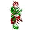


 PDBj
PDBj

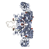
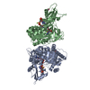
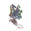
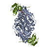
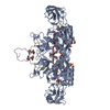
 Assembly


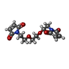

 Mass: 427.201 Da / Num. of mol.: 1 / Source method: obtained synthetically / Formula: C10H15N5O10P2 / Comment: ADP, energy-carrying molecule*YM
 Mass: 24.305 Da / Num. of mol.: 2 / Source method: obtained synthetically / Formula: Mg
 Mass: 118.174 Da / Num. of mol.: 3 / Source method: obtained synthetically / Formula: C6H14O2 / Comment: precipitant*YM
 Mass: 59.044 Da / Num. of mol.: 8 / Source method: obtained synthetically / Formula: C2H3O2
 Mass: 94.971 Da / Num. of mol.: 1 / Source method: obtained synthetically / Formula: PO4
 Mass: 22.990 Da / Num. of mol.: 1 / Source method: obtained synthetically / Formula: Na
 Mass: 308.287 Da / Num. of mol.: 1 / Source method: obtained synthetically / Formula: C14H16N2O6
 Sample preparation
 / Beamline: MASSIF-3 / Wavelength: 0.9677 Å
 Processing