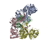
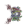
Entry Database : PDB / ID : 4i7zTitle Crystal structure of cytochrome b6f in DOPG, with disordered Rieske Iron-Sulfur Protein soluble domain (Cytochrome b6-f complex subunit ...) x 5 Apocytochrome f Cytochrome b6 Cytochrome b6-f complex iron-sulfur subunit Keywords / / / / / Function / homology Function Domain/homology Component
/ / / / / / / / / / / / / / / / / / / / / / / / / / / / / / / / / / / / / / / / / / / / / / / / / / / / / / / / / / / / / / / / / / / / / / / / / / / / / / / / / / / / / / / / / / / / / / / / / / / / / / / / / / / / / / / / / / Biological species Mastigocladus laminosus (bacteria)Method / / Resolution : 2.803 Å Authors Hasan, S.S. / Stofleth, J.T. / Yamashita, E. / Cramer, W.A. Journal : Biochemistry / Year : 2013Title : Lipid-induced conformational changes within the cytochrome b6f complex of oxygenic photosynthesis.Authors : Hasan, S.S. / Stofleth, J.T. / Yamashita, E. / Cramer, W.A. History Deposition Dec 1, 2012 Deposition site / Processing site Revision 1.0 Apr 17, 2013 Provider / Type Revision 1.1 Dec 4, 2013 Group Revision 1.2 Nov 15, 2017 Group / Category / Item Revision 1.3 Jul 17, 2019 Group / Refinement description / Category Item / _software.name / _software.versionRevision 2.0 Feb 28, 2024 Group Data collection / Database references ... Data collection / Database references / Derived calculations / Non-polymer description Category chem_comp / chem_comp_atom ... chem_comp / chem_comp_atom / chem_comp_bond / database_2 / pdbx_struct_conn_angle / pdbx_validate_chiral / struct_conn / struct_ref_seq_dif / struct_site Item _chem_comp.formula / _database_2.pdbx_DOI ... _chem_comp.formula / _database_2.pdbx_DOI / _database_2.pdbx_database_accession / _pdbx_struct_conn_angle.ptnr1_auth_asym_id / _pdbx_struct_conn_angle.ptnr1_auth_comp_id / _pdbx_struct_conn_angle.ptnr1_auth_seq_id / _pdbx_struct_conn_angle.ptnr1_label_asym_id / _pdbx_struct_conn_angle.ptnr1_label_atom_id / _pdbx_struct_conn_angle.ptnr1_label_comp_id / _pdbx_struct_conn_angle.ptnr1_label_seq_id / _pdbx_struct_conn_angle.ptnr2_auth_asym_id / _pdbx_struct_conn_angle.ptnr2_auth_comp_id / _pdbx_struct_conn_angle.ptnr2_auth_seq_id / _pdbx_struct_conn_angle.ptnr2_label_asym_id / _pdbx_struct_conn_angle.ptnr2_label_atom_id / _pdbx_struct_conn_angle.ptnr2_label_comp_id / _pdbx_struct_conn_angle.ptnr3_auth_asym_id / _pdbx_struct_conn_angle.ptnr3_auth_comp_id / _pdbx_struct_conn_angle.ptnr3_auth_seq_id / _pdbx_struct_conn_angle.ptnr3_label_asym_id / _pdbx_struct_conn_angle.ptnr3_label_atom_id / _pdbx_struct_conn_angle.ptnr3_label_comp_id / _pdbx_struct_conn_angle.ptnr3_label_seq_id / _pdbx_struct_conn_angle.value / _struct_conn.pdbx_dist_value / _struct_conn.ptnr1_auth_asym_id / _struct_conn.ptnr1_auth_comp_id / _struct_conn.ptnr1_auth_seq_id / _struct_conn.ptnr1_label_asym_id / _struct_conn.ptnr1_label_atom_id / _struct_conn.ptnr1_label_comp_id / _struct_conn.ptnr1_label_seq_id / _struct_conn.ptnr2_auth_asym_id / _struct_conn.ptnr2_auth_comp_id / _struct_conn.ptnr2_auth_seq_id / _struct_conn.ptnr2_label_asym_id / _struct_conn.ptnr2_label_atom_id / _struct_conn.ptnr2_label_comp_id / _struct_conn.ptnr2_label_seq_id / _struct_ref_seq_dif.details / _struct_site.pdbx_auth_asym_id / _struct_site.pdbx_auth_comp_id / _struct_site.pdbx_auth_seq_id
Show all Show less
 Movie
Movie Controller

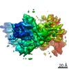
 Yorodumi
 Open data
 Basic information
 Components
 Keywords
 Function and homology information
 Mastigocladus laminosus (bacteria)
 X-RAY DIFFRACTION /
  Authors
 Citation
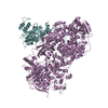
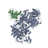
 Structure visualization
 Downloads & links
 Other downloads
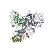
 PDBj
PDBj


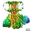
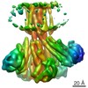
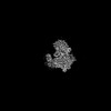
 Assembly
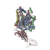


 Mass: 616.487 Da / Num. of mol.: 4 / Source method: obtained synthetically / Formula: C34H32FeN4O4
 Mass: 212.415 Da / Num. of mol.: 1 / Source method: obtained synthetically / Formula: C15H32
 Mass: 254.494 Da / Num. of mol.: 1 / Source method: obtained synthetically / Formula: C18H38
 Mass: 496.589 Da / Num. of mol.: 3 / Source method: obtained synthetically / Formula: C23H44O11 / Comment: detergent*YM
 Mass: 112.411 Da / Num. of mol.: 3 / Source method: obtained synthetically / Formula: Cd
 Mass: 893.489 Da / Num. of mol.: 1 / Source method: obtained synthetically / Formula: C55H72MgN4O5
 Mass: 704.912 Da / Num. of mol.: 3 / Source method: obtained synthetically / Formula: C37H69O10P
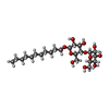
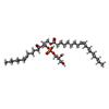
 Mass: 360.335 Da / Num. of mol.: 1 / Source method: obtained synthetically / Formula: C11H20O11S
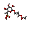
 Mass: 114.229 Da / Num. of mol.: 1 / Source method: obtained synthetically / Formula: C8H18
 Mass: 536.873 Da / Num. of mol.: 1 / Source method: obtained synthetically / Formula: C40H56
 Sample preparation
 Processing