 Movie
Movie Controller

[English] 日本語
 Yorodumi
- PDB-6hee: Crystal structure of Extracellular Domain 1 (ECD1) of FtsX from S... -
+ Open data

- Basic information
| Entry | Database: PDB / ID: 6hee | ||||||
|---|---|---|---|---|---|---|---|
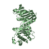
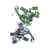
| Title | Crystal structure of Extracellular Domain 1 (ECD1) of FtsX from S. pneumonie in complex with undecyl-maltoside | ||||||
 Components Components | Cell division protein FtsX | ||||||
 Keywords Keywords | CELL CYCLE / cell division / MEMBRANE PROTEIN | ||||||
| Function / homology |  Function and homology information Function and homology information | ||||||
| Biological species | Streptococcus pneumoniae serotype 2 | ||||||
| Method |  X-RAY DIFFRACTION / SYNCHROTRON / Resolution: 2.3 Å X-RAY DIFFRACTION / SYNCHROTRON / Resolution: 2.3 Å | ||||||
 Authors Authors | Martinez-Caballero, S. / Alcorlo-Pages, M. / Hermoso, J.A. | ||||||
 Citation Citation | Journal: Mbio / Year: 2019 Title: Structure of the Large Extracellular Loop of FtsX and Its Interaction with the Essential Peptidoglycan Hydrolase PcsB in Streptococcus pneumoniae. Authors: Rued, B.E. / Alcorlo, M. / Edmonds, K.A. / Martinez-Caballero, S. / Straume, D. / Fu, Y. / Bruce, K.E. / Wu, H. / Havarstein, L.S. / Hermoso, J.A. / Winkler, M.E. / Giedroc, D.P. | ||||||
| History |
|
- Structure visualization
| Structure viewer | Molecule: MolmilJmol/JSmol |
|---|
- Downloads & links
-Download
| PDBx/mmCIF format | 6hee.cif.gz | 59.6 KB | Display | PDBx/mmCIF format |
|---|---|---|---|---|
| PDB format | pdb6hee.ent.gz | 42.1 KB | Display | PDB format |
| PDBx/mmJSON format | 6hee.json.gz | Tree view | PDBx/mmJSON format | |
| Others |  Other downloads Other downloads |
-Validation report
| Arichive directory | https://data.pdbj.org/pub/pdb/validation_reports/he/6heeftp://data.pdbj.org/pub/pdb/validation_reports/he/6hee | HTTPS FTP |
|---|
-Related structure data
| Related structure data | 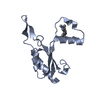 6he6SC  6hfxC 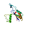 6mk7C S: Starting model for refinement C: citing same article ( |
|---|---|
| Similar structure data |


-Links
 PDBj
PDBj- Assembly
| Deposited unit | 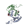
| ||||||||
|---|---|---|---|---|---|---|---|---|---|
| 1 | 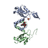
| ||||||||
| 2 |
| ||||||||
| Unit cell |
|
-Components
| #1: Protein | Mass: 13325.637 Da / Num. of mol.: 2 Source method: isolated from a genetically manipulated source Source: (gene. exp.)  Streptococcus pneumoniae serotype 2 (strain D39 / NCTC 7466) (bacteria) Streptococcus pneumoniae serotype 2 (strain D39 / NCTC 7466) (bacteria)Strain: D39 / NCTC 7466 / Gene: ftsX, SPD_0660 / Production host: #2: Chemical | ChemComp-UMQ / |   Mass: 496.589 Da / Num. of mol.: 1 / Source method: obtained synthetically / Formula: C23H44O11 / Comment: detergent*YM Mass: 496.589 Da / Num. of mol.: 1 / Source method: obtained synthetically / Formula: C23H44O11 / Comment: detergent*YM#3: Chemical | ChemComp-SO4 / |   Mass: 96.063 Da / Num. of mol.: 1 / Source method: obtained synthetically / Formula: SO4 Mass: 96.063 Da / Num. of mol.: 1 / Source method: obtained synthetically / Formula: SO4#4: Chemical | ChemComp-TRS / |   Mass: 122.143 Da / Num. of mol.: 1 / Source method: obtained synthetically / Formula: C4H12NO3 / Comment: pH buffer*YM Mass: 122.143 Da / Num. of mol.: 1 / Source method: obtained synthetically / Formula: C4H12NO3 / Comment: pH buffer*YM#5: Water | ChemComp-HOH / |  Mass: 18.015 Da / Num. of mol.: 21 / Source method: isolated from a natural source / Formula: H2O Mass: 18.015 Da / Num. of mol.: 21 / Source method: isolated from a natural source / Formula: H2O |
|---|
-Experimental details
-Experiment
| Experiment | Method: X-RAY DIFFRACTION / Number of used crystals: 1 |
|---|
- Sample preparation
| Crystal | Density Matthews: 3.57 Å3/Da / Density % sol: 65.5 % |
|---|---|
| Crystal grow | Temperature: 291.15 K / Method: vapor diffusion, sitting drop Details: ammonium sulfate, sodium and potassium tartrate and sodium citrate |
-Data collection
| Diffraction | Mean temperature: 100 K |
|---|---|
| Diffraction source | Source: SYNCHROTRON / Site: ALBA  / Beamline: XALOC / Wavelength: 0.97934 Å / Beamline: XALOC / Wavelength: 0.97934 Å |
| Detector | Type: DECTRIS PILATUS 6M / Detector: PIXEL / Date: Sep 13, 2016 |
| Radiation | Protocol: SINGLE WAVELENGTH / Monochromatic (M) / Laue (L): M / Scattering type: x-ray |
| Radiation wavelength | Wavelength: 0.97934 Å / Relative weight: 1 |
| Reflection | Resolution: 2.3→42.31 Å / Num. obs: 17731 / % possible obs: 98.89 % / Redundancy: 22.1 % / Rpim(I) all: 0.004 / Net I/σ(I): 44.14 |
| Reflection shell | Resolution: 2.3→2.38 Å / Rpim(I) all: 0.092 |
- Processing
| Software |
| ||||||||||||||||||||||||||||||||||||||||||||||||||||||||
|---|---|---|---|---|---|---|---|---|---|---|---|---|---|---|---|---|---|---|---|---|---|---|---|---|---|---|---|---|---|---|---|---|---|---|---|---|---|---|---|---|---|---|---|---|---|---|---|---|---|---|---|---|---|---|---|---|---|
| Refinement | Starting model: 6HE6 Resolution: 2.3→42.31 Å / SU ML: 0.34 / Cross valid method: THROUGHOUT / σ(F): 1.34 / Phase error: 34.18
| ||||||||||||||||||||||||||||||||||||||||||||||||||||||||
| Solvent computation | Shrinkage radii: 0.9 Å / VDW probe radii: 1.11 Å | ||||||||||||||||||||||||||||||||||||||||||||||||||||||||
| Refinement step | Cycle: LAST / Resolution: 2.3→42.31 Å
| ||||||||||||||||||||||||||||||||||||||||||||||||||||||||
| Refine LS restraints |
| ||||||||||||||||||||||||||||||||||||||||||||||||||||||||
| LS refinement shell |
|
