 Movie
Movie Controller

[English] 日本語
 Yorodumi
- PDB-6hdm: R49A variant of beta-phosphoglucomutase from Lactococcus lactis c... -
+ Open data

- Basic information
| Entry | Database: PDB / ID: 6hdm | |||||||||
|---|---|---|---|---|---|---|---|---|---|---|
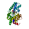
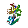
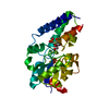
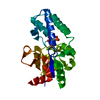
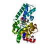
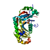
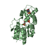
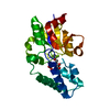
| Title | R49A variant of beta-phosphoglucomutase from Lactococcus lactis complexed with magnesium trifluoride and beta-G6P to 1.3 A. | |||||||||
 Components Components | Beta-phosphoglucomutase | |||||||||
 Keywords Keywords | ISOMERASE / phosphoglucomutase / metal fluoride / transition state analog | |||||||||
| Function / homology |  Function and homology information Function and homology informationbeta-phosphoglucomutase / beta-phosphoglucomutase activity / carbohydrate metabolic process / magnesium ion binding / cytoplasm Similarity search - Function | |||||||||
| Biological species |  Lactococcus lactis subsp. lactis (lactic acid bacteria) Lactococcus lactis subsp. lactis (lactic acid bacteria) | |||||||||
| Method |  X-RAY DIFFRACTION / SYNCHROTRON / MOLECULAR REPLACEMENT / Resolution: 1.3 Å X-RAY DIFFRACTION / SYNCHROTRON / MOLECULAR REPLACEMENT / Resolution: 1.3 Å | |||||||||
 Authors Authors | Robertson, A.J. / Bisson, C. / Waltho, J.P. | |||||||||
| Funding support |  United Kingdom, 2items United Kingdom, 2items
| |||||||||
 Citation Citation | Journal: To Be Published Title: Transition state of phospho-enzyme hydrolysis in beta-phosphoglucomutase. Authors: Robertson, A.J. / Bisson, C. / Waltho, J.P. | |||||||||
| History |
|
- Structure visualization
| Structure viewer | Molecule: MolmilJmol/JSmol |
|---|
- Downloads & links
-Download
| PDBx/mmCIF format | 6hdm.cif.gz | 114.5 KB | Display | PDBx/mmCIF format |
|---|---|---|---|---|
| PDB format | pdb6hdm.ent.gz | 87.2 KB | Display | PDB format |
| PDBx/mmJSON format | 6hdm.json.gz | Tree view | PDBx/mmJSON format | |
| Others |  Other downloads Other downloads |
-Validation report
| Arichive directory | https://data.pdbj.org/pub/pdb/validation_reports/hd/6hdmftp://data.pdbj.org/pub/pdb/validation_reports/hd/6hdm | HTTPS FTP |
|---|
-Related structure data
| Related structure data | 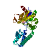 6h8uC  6h8vC 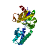 6h8wC  6h8xC 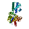 6h8yC 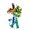 6h8zC 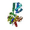 6h90C 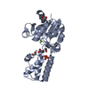 6h93C 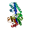 6h94C 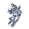 6hdfC 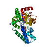 6hdgC 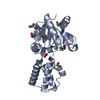 6hdhC  6hdiC  6hdjC  6hdkC  6hdlC  2wf5S S: Starting model for refinement C: citing same article ( |
|---|---|
| Similar structure data |
-Links
 PDBj
PDBj

- Assembly
| Deposited unit | 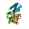
| ||||||||
|---|---|---|---|---|---|---|---|---|---|
| 1 |
| ||||||||
| Unit cell |
|
-Components
-Protein / Sugars , 2 types, 2 molecules A
| #1: Protein | Mass: 24153.479 Da / Num. of mol.: 1 / Mutation: K125R, Y206H, R49A Source method: isolated from a genetically manipulated source Source: (gene. exp.) Lactococcus lactis subsp. lactis (lactic acid bacteria)Strain: IL1403 / Gene: pgmB, LL0429, L0001 / Production host: |
|---|---|
| #5: Sugar | ChemComp-BG6 /  Type: D-saccharide, beta linking / Mass: 260.136 Da / Num. of mol.: 1 Type: D-saccharide, beta linking / Mass: 260.136 Da / Num. of mol.: 1Source method: isolated from a genetically manipulated source Formula: C6H13O9P / Feature type: SUBJECT OF INVESTIGATION |
-Non-polymers , 6 types, 213 molecules 


| #2: Chemical | ChemComp-MG /  Mass: 24.305 Da / Num. of mol.: 1 / Source method: obtained synthetically / Formula: Mg Mass: 24.305 Da / Num. of mol.: 1 / Source method: obtained synthetically / Formula: Mg | ||
|---|---|---|---|
| #3: Chemical | ChemComp-MGF /  Mass: 81.300 Da / Num. of mol.: 1 / Source method: obtained synthetically / Formula: F3Mg / Feature type: SUBJECT OF INVESTIGATION Mass: 81.300 Da / Num. of mol.: 1 / Source method: obtained synthetically / Formula: F3Mg / Feature type: SUBJECT OF INVESTIGATION | ||
| #4: Chemical | ChemComp-NA /  Mass: 22.990 Da / Num. of mol.: 1 / Source method: obtained synthetically / Formula: Na Mass: 22.990 Da / Num. of mol.: 1 / Source method: obtained synthetically / Formula: Na | ||
| #6: Chemical | ChemComp-PDO /  Mass: 76.094 Da / Num. of mol.: 1 / Source method: obtained synthetically / Formula: C3H8O2 Mass: 76.094 Da / Num. of mol.: 1 / Source method: obtained synthetically / Formula: C3H8O2 | ||
| #7: Chemical |  Mass: 62.068 Da / Num. of mol.: 2 / Source method: obtained synthetically / Formula: C2H6O2 Mass: 62.068 Da / Num. of mol.: 2 / Source method: obtained synthetically / Formula: C2H6O2#8: Water | ChemComp-HOH / | Mass: 18.015 Da / Num. of mol.: 207 / Source method: isolated from a natural source / Formula: H2O |
-Experimental details
-Experiment
| Experiment | Method: X-RAY DIFFRACTION / Number of used crystals: 1 |
|---|
- Sample preparation
| Crystal | Density Matthews: 2.19 Å3/Da / Density % sol: 43.96 % |
|---|---|
| Crystal grow | Temperature: 291 K / Method: vapor diffusion, hanging drop / pH: 7.2 Details: 24-34% PEG 4000 200mM sodium acetate 50mM TRIS pH 7.5 |
-Data collection
| Diffraction | Mean temperature: 100 K |
|---|---|
| Diffraction source | Source: SYNCHROTRON / Site: Diamond / Beamline: I03 / Wavelength: 0.97625 Å |
| Detector | Type: DECTRIS PILATUS3 S 6M / Detector: PIXEL / Date: Oct 9, 2017 |
| Radiation | Protocol: SINGLE WAVELENGTH / Monochromatic (M) / Laue (L): M / Scattering type: x-ray |
| Radiation wavelength | Wavelength: 0.97625 Å / Relative weight: 1 |
| Reflection | Resolution: 1.3→54.34 Å / Num. obs: 53048 / % possible obs: 99.7 % / Redundancy: 6.8 % / CC1/2: 0.999 / Rmerge(I) obs: 0.052 / Rpim(I) all: 0.022 / Rrim(I) all: 0.057 / Net I/σ(I): 21.1 |
| Reflection shell | Resolution: 1.3→1.32 Å / Redundancy: 4.5 % / Rmerge(I) obs: 0.263 / Num. unique obs: 2526 / CC1/2: 0.944 / Rpim(I) all: 0.138 / Rrim(I) all: 0.299 / % possible all: 95.3 |
- Processing
| Software |
| ||||||||||||||||||||||||||||||||||||||||||||||||||||||||||||||||||||||||||||||||||||||||||||||||||||||||||||||||||||||||||||||||||||||||||||||||||||||||||||||||||||||||||||||||||||||
|---|---|---|---|---|---|---|---|---|---|---|---|---|---|---|---|---|---|---|---|---|---|---|---|---|---|---|---|---|---|---|---|---|---|---|---|---|---|---|---|---|---|---|---|---|---|---|---|---|---|---|---|---|---|---|---|---|---|---|---|---|---|---|---|---|---|---|---|---|---|---|---|---|---|---|---|---|---|---|---|---|---|---|---|---|---|---|---|---|---|---|---|---|---|---|---|---|---|---|---|---|---|---|---|---|---|---|---|---|---|---|---|---|---|---|---|---|---|---|---|---|---|---|---|---|---|---|---|---|---|---|---|---|---|---|---|---|---|---|---|---|---|---|---|---|---|---|---|---|---|---|---|---|---|---|---|---|---|---|---|---|---|---|---|---|---|---|---|---|---|---|---|---|---|---|---|---|---|---|---|---|---|---|---|
| Refinement | Method to determine structure: MOLECULAR REPLACEMENT Starting model: 2WF5 Resolution: 1.3→48.27 Å / Cor.coef. Fo:Fc: 0.976 / Cor.coef. Fo:Fc free: 0.972 / SU B: 1.114 / SU ML: 0.022 / Cross valid method: THROUGHOUT / ESU R: 0.042 / ESU R Free: 0.04 / Stereochemistry target values: MAXIMUM LIKELIHOOD / Details: HYDROGENS HAVE BEEN ADDED IN THE RIDING POSITIONS
| ||||||||||||||||||||||||||||||||||||||||||||||||||||||||||||||||||||||||||||||||||||||||||||||||||||||||||||||||||||||||||||||||||||||||||||||||||||||||||||||||||||||||||||||||||||||
| Solvent computation | Ion probe radii: 0.8 Å / Shrinkage radii: 0.8 Å / VDW probe radii: 1.2 Å / Solvent model: MASK | ||||||||||||||||||||||||||||||||||||||||||||||||||||||||||||||||||||||||||||||||||||||||||||||||||||||||||||||||||||||||||||||||||||||||||||||||||||||||||||||||||||||||||||||||||||||
| Displacement parameters | Biso mean: 15.053 Å2
| ||||||||||||||||||||||||||||||||||||||||||||||||||||||||||||||||||||||||||||||||||||||||||||||||||||||||||||||||||||||||||||||||||||||||||||||||||||||||||||||||||||||||||||||||||||||
| Refinement step | Cycle: 1 / Resolution: 1.3→48.27 Å
| ||||||||||||||||||||||||||||||||||||||||||||||||||||||||||||||||||||||||||||||||||||||||||||||||||||||||||||||||||||||||||||||||||||||||||||||||||||||||||||||||||||||||||||||||||||||
| Refine LS restraints |
|