 Movie
Movie Controller


+ Open data

- Basic information
| Entry | Database: PDB / ID: 6hdd | |||||||||
|---|---|---|---|---|---|---|---|---|---|---|
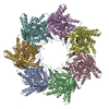
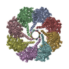
| Title | OBP chaperonin in the nucleotide-free state | |||||||||
 Components Components | Putative chaperonin GroEL | |||||||||
 Keywords Keywords | CHAPERONE / chaperonin / nucleotide-free | |||||||||
| Function / homology |  Function and homology information Function and homology informationATP-dependent protein folding chaperone / protein refolding / ATP binding Similarity search - Function | |||||||||
| Biological species |  Pseudomonas phage OBP (virus) Pseudomonas phage OBP (virus) | |||||||||
| Method | ELECTRON MICROSCOPY / single particle reconstruction / cryo EM / Resolution: 4.5 Å | |||||||||
 Authors Authors | Semenyuk, P.I. / Stanishneva-Konovalova, T.B. / Sokolova, O.S. | |||||||||
| Funding support |  Russian Federation, 2items Russian Federation, 2items
| |||||||||
 Citation Citation | Journal: J Struct Biol / Year: 2020 Title: Cryo-EM reveals an asymmetry in a novel single-ring viral chaperonin. Authors: Tatiana B Stanishneva-Konovalova / Pavel I Semenyuk / Lidia P Kurochkina / Evgeny B Pichkur / Alexander L Vasilyev / Mikhail V Kovalchuk / Mikhail P Kirpichnikov / Olga S Sokolova / Abstract: Chaperonins are ubiquitously present protein complexes, which assist the proper folding of newly synthesized proteins and prevent aggregation of denatured proteins in an ATP-dependent manner. They ...Chaperonins are ubiquitously present protein complexes, which assist the proper folding of newly synthesized proteins and prevent aggregation of denatured proteins in an ATP-dependent manner. They are classified into group I (bacterial, mitochondrial, chloroplast chaperonins) and group II (archaeal and eukaryotic cytosolic variants). However, both of these groups do not include recently discovered viral chaperonins. Here, we solved the symmetry-free cryo-EM structures of a single-ring chaperonin encoded by the gene 246 of bacteriophage OBP Pseudomonas fluorescens, in the nucleotide-free, ATPγS-, and ADP-bound states, with resolutions of 4.3 Å, 5.0 Å, and 6 Å, respectively. The structure of OBP chaperonin reveals a unique subunit arrangement, with three pairs of subunits and one unpaired subunit. Each pair combines subunits in two possible conformations, differing in nucleotide-binding affinity. The binding of nucleotides results in the increase of subunits' conformational variability. Due to its unique structural and functional features, OBP chaperonin can represent a new group. | |||||||||
| History |
|
- Structure visualization
| Movie |
Movie viewer |
|---|---|
| Structure viewer | Molecule: MolmilJmol/JSmol |
- Downloads & links
-Download
| PDBx/mmCIF format | 6hdd.cif.gz | 600.2 KB | Display | PDBx/mmCIF format |
|---|---|---|---|---|
| PDB format | pdb6hdd.ent.gz | 504 KB | Display | PDB format |
| PDBx/mmJSON format | 6hdd.json.gz | Tree view | PDBx/mmJSON format | |
| Others |  Other downloads Other downloads |
-Validation report
| Summary document | 6hdd_validation.pdf.gz | 953.6 KB | Display | wwPDB validaton report |
|---|---|---|---|---|
| Full document | 6hdd_full_validation.pdf.gz | 1014.2 KB | Display | |
| Data in XML | 6hdd_validation.xml.gz | 102.8 KB | Display | |
| Data in CIF | 6hdd_validation.cif.gz | 154.4 KB | Display | |
| Arichive directory | https://data.pdbj.org/pub/pdb/validation_reports/hd/6hddftp://data.pdbj.org/pub/pdb/validation_reports/hd/6hdd | HTTPS FTP |
-Related structure data
| Related structure data |  0204MC  0191C  0208C C: citing same article ( M: map data used to model this data |
|---|---|
| Similar structure data |


-Links
 PDBj
PDBj
- Assembly
| Deposited unit | 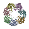
|
|---|---|
| 1 |
|
-Components
| #1: Protein | Mass: 57259.703 Da / Num. of mol.: 7 Source method: isolated from a genetically manipulated source Source: (gene. exp.) Pseudomonas phage OBP (virus) / Gene: OBP_246 / Production host:  |
|---|
-Experimental details
-Experiment
| Experiment | Method: ELECTRON MICROSCOPY |
|---|---|
| EM experiment | Aggregation state: PARTICLE / 3D reconstruction method: single particle reconstruction |
- Sample preparation
| Component | Name: OBP chaperonin in the nucleotide-free state / Type: COMPLEX / Entity ID: all / Source: RECOMBINANT |
|---|---|
| Molecular weight | Value: 0.42 MDa / Experimental value: YES |
| Source (natural) | Organism: Pseudomonas phage OBP (virus) |
| Source (recombinant) | Organism: |
| Buffer solution | pH: 7.5 |
| Specimen | Conc.: 2.5 mg/ml / Embedding applied: NO / Shadowing applied: NO / Staining applied: NO / Vitrification applied: YES |
| Vitrification | Cryogen name: ETHANE |
- Electron microscopy imaging
| Experimental equipment |  Model: Titan Krios / Image courtesy: FEI Company |
|---|---|
| Microscopy | Model: FEI TITAN KRIOS |
| Electron gun | Electron source:  FIELD EMISSION GUN / Accelerating voltage: 300 kV / Illumination mode: FLOOD BEAM FIELD EMISSION GUN / Accelerating voltage: 300 kV / Illumination mode: FLOOD BEAM |
| Electron lens | Mode: BRIGHT FIELD |
| Image recording | Electron dose: 100 e/Å2 / Film or detector model: FEI FALCON II (4k x 4k) |
- Processing
| CTF correction | Type: PHASE FLIPPING AND AMPLITUDE CORRECTION |
|---|---|
| Symmetry | Point symmetry: C1 (asymmetric) |
| 3D reconstruction | Resolution: 4.5 Å / Resolution method: FSC 0.143 CUT-OFF / Num. of particles: 127180 / Symmetry type: POINT |
