 Movie
Movie Controller

[English] 日本語
 Yorodumi
- PDB-6hcx: Influenza Virus N9 Neuraminidase A complex with Zanamivir molecul... -
+ Open data

- Basic information
| Entry | Database: PDB / ID: 6hcx | |||||||||
|---|---|---|---|---|---|---|---|---|---|---|
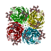
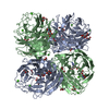
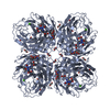
| Title | Influenza Virus N9 Neuraminidase A complex with Zanamivir molecule (Tern). | |||||||||
 Components Components | Neuraminidase | |||||||||
 Keywords Keywords | HYDROLASE / Complex / Zanamivir / Relenza / Neuraminidase / N9 / Influenza / Virus / Enzyme / Inhibitor | |||||||||
| Function / homology |  Function and homology information Function and homology informationexo-alpha-sialidase / exo-alpha-sialidase activity / viral budding from plasma membrane / carbohydrate metabolic process / host cell plasma membrane / virion membrane / metal ion binding / membrane Similarity search - Function | |||||||||
| Biological species |   Influenza A virus Influenza A virus | |||||||||
| Method |  X-RAY DIFFRACTION / SYNCHROTRON / MOLECULAR REPLACEMENT / Resolution: 1.3 Å X-RAY DIFFRACTION / SYNCHROTRON / MOLECULAR REPLACEMENT / Resolution: 1.3 Å | |||||||||
 Authors Authors | Salinger, M.T. / Hobbs, J.R. / Murray, J.W. / Laver, W.G. / Kuhn, P. / Garman, E.F. | |||||||||
| Funding support |  United Kingdom, 1items United Kingdom, 1items
| |||||||||
 Citation Citation | Journal: To Be Published Title: High Resolution Structures of Viral Neuraminidase with Drugs Bound in the Active Site. (In preparation) Authors: Salinger, M.T. / Hobbs, J.R. / Murray, J.W. / Laver, W.G. / Kuhn, P. / Garman, E.F. | |||||||||
| History |
|
- Structure visualization
| Structure viewer | Molecule: MolmilJmol/JSmol |
|---|
- Downloads & links
-Download
| PDBx/mmCIF format | 6hcx.cif.gz | 216.5 KB | Display | PDBx/mmCIF format |
|---|---|---|---|---|
| PDB format | pdb6hcx.ent.gz | 170.3 KB | Display | PDB format |
| PDBx/mmJSON format | 6hcx.json.gz | Tree view | PDBx/mmJSON format | |
| Others |  Other downloads Other downloads |
-Validation report
| Summary document | 6hcx_validation.pdf.gz | 1.8 MB | Display | wwPDB validaton report |
|---|---|---|---|---|
| Full document | 6hcx_full_validation.pdf.gz | 1.8 MB | Display | |
| Data in XML | 6hcx_validation.xml.gz | 25.5 KB | Display | |
| Data in CIF | 6hcx_validation.cif.gz | 40.1 KB | Display | |
| Arichive directory | https://data.pdbj.org/pub/pdb/validation_reports/hc/6hcxftp://data.pdbj.org/pub/pdb/validation_reports/hc/6hcx | HTTPS FTP |
-Related structure data
| Related structure data |  6hfcC  6hfyC  6hg0C  6hgbC 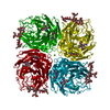 1f8eS S: Starting model for refinement C: citing same article ( |
|---|---|
| Similar structure data |


-Links
 PDBj
PDBj

- Assembly
| Deposited unit | 
| ||||||||||||||||||
|---|---|---|---|---|---|---|---|---|---|---|---|---|---|---|---|---|---|---|---|
| 1 | 
| ||||||||||||||||||
| Unit cell |
| ||||||||||||||||||
| Components on special symmetry positions |
|
-Components
-Protein , 1 types, 1 molecules A
| #1: Protein | Mass: 43723.770 Da / Num. of mol.: 1 Source method: isolated from a genetically manipulated source Source: (gene. exp.) Influenza A virus (strain A/Tern/Australia/G70C/1975 H11N9)Gene: NA / Production host:  |
|---|
-Sugars , 3 types, 4 molecules 
| #2: Polysaccharide | alpha-D-mannopyranose-(1-2)-alpha-D-mannopyranose-(1-2)-alpha-D-mannopyranose-(1-3)-[alpha-D- ...alpha-D-mannopyranose-(1-2)-alpha-D-mannopyranose-(1-2)-alpha-D-mannopyranose-(1-3)-[alpha-D-mannopyranose-(1-3)-[alpha-D-mannopyranose-(1-6)]alpha-D-mannopyranose-(1-6)]beta-D-mannopyranose-(1-4)-2-acetamido-2-deoxy-beta-D-glucopyranose-(1-4)-2-acetamido-2-deoxy-beta-D-glucopyranose Source method: isolated from a genetically manipulated source | ||
|---|---|---|---|
| #3: Polysaccharide | Source method: isolated from a genetically manipulated source #6: Sugar | ChemComp-ZMR / |  Type: D-saccharide / Mass: 332.310 Da / Num. of mol.: 1 Type: D-saccharide / Mass: 332.310 Da / Num. of mol.: 1Source method: isolated from a genetically manipulated source Formula: C12H20N4O7 / Comment: medication*YM |
-Non-polymers , 4 types, 697 molecules 


| #4: Chemical |  Mass: 40.078 Da / Num. of mol.: 2 Mass: 40.078 Da / Num. of mol.: 2Source method: isolated from a genetically manipulated source Formula: Ca #5: Chemical |  Mass: 92.094 Da / Num. of mol.: 3 Mass: 92.094 Da / Num. of mol.: 3Source method: isolated from a genetically manipulated source Formula: C3H8O3 #7: Chemical | ChemComp-K / |  Mass: 39.098 Da / Num. of mol.: 1 Mass: 39.098 Da / Num. of mol.: 1Source method: isolated from a genetically manipulated source Formula: K #8: Water | ChemComp-HOH / | Mass: 18.015 Da / Num. of mol.: 691 / Source method: isolated from a natural source / Formula: H2O |
|---|
-Details
| Has protein modification | Y |
|---|
-Experimental details
-Experiment
| Experiment | Method: X-RAY DIFFRACTION / Number of used crystals: 1 |
|---|
- Sample preparation
| Crystal | Density Matthews: 2.82 Å3/Da / Density % sol: 56.42 % |
|---|---|
| Crystal grow | Temperature: 293 K / Method: vapor diffusion, hanging drop / pH: 6.6 Details: N9 crystals were grown by hanging-drop vapour diffusion against a reservoir of 1.9M potassium phosphate, pH 6.8, starting with equal volumes of N9 NA (10-15 mg/ml in water) and potassium ...Details: N9 crystals were grown by hanging-drop vapour diffusion against a reservoir of 1.9M potassium phosphate, pH 6.8, starting with equal volumes of N9 NA (10-15 mg/ml in water) and potassium phosphate buffer 1.4M KH2PO4:3M K2HPO4 in ratio 8:4, pH 6.6 at 20 degrees celsius. Inhibitor complexes obtained by soaking N9 crystals in a solution of 1.4M potassium phosphate buffer, pH 6.8, containing 5mM of inhibitor for 3 hours at 18 degrees celsius. Soaked in glycerol cryo-buffer. |
-Data collection
| Diffraction | Mean temperature: 100 K |
|---|---|
| Diffraction source | Source: SYNCHROTRON / Site: SSRL  / Beamline: BL11-1 / Wavelength: 0.98 Å / Beamline: BL11-1 / Wavelength: 0.98 Å |
| Detector | Type: ADSC QUANTUM 315 / Detector: CCD / Date: Dec 12, 2001 |
| Radiation | Protocol: SINGLE WAVELENGTH / Monochromatic (M) / Laue (L): M / Scattering type: x-ray |
| Radiation wavelength | Wavelength: 0.98 Å / Relative weight: 1 |
| Reflection | Resolution: 1.29→42.68 Å / Num. obs: 122689 / % possible obs: 99.9 % / Redundancy: 10 % / Biso Wilson estimate: 12.3 Å2 / Rrim(I) all: 0.093 / Net I/σ(I): 16.3 |
| Reflection shell | Resolution: 1.3→1.4 Å / Redundancy: 6.3 % / Mean I/σ(I) obs: 2.3 / Num. unique obs: 8993 / Rrim(I) all: 0.706 / % possible all: 99.5 |
- Processing
| Software |
| ||||||||||||||||||||||||||||||||||||||||||||||||||||||||||||||||||||||||||||||||||||||||||||||||||||||||||||||||||||||||||||||||||||||||||||||||||||||||||||||||||||||||||||||||||||||
|---|---|---|---|---|---|---|---|---|---|---|---|---|---|---|---|---|---|---|---|---|---|---|---|---|---|---|---|---|---|---|---|---|---|---|---|---|---|---|---|---|---|---|---|---|---|---|---|---|---|---|---|---|---|---|---|---|---|---|---|---|---|---|---|---|---|---|---|---|---|---|---|---|---|---|---|---|---|---|---|---|---|---|---|---|---|---|---|---|---|---|---|---|---|---|---|---|---|---|---|---|---|---|---|---|---|---|---|---|---|---|---|---|---|---|---|---|---|---|---|---|---|---|---|---|---|---|---|---|---|---|---|---|---|---|---|---|---|---|---|---|---|---|---|---|---|---|---|---|---|---|---|---|---|---|---|---|---|---|---|---|---|---|---|---|---|---|---|---|---|---|---|---|---|---|---|---|---|---|---|---|---|---|---|
| Refinement | Method to determine structure: MOLECULAR REPLACEMENT Starting model: 1F8E Resolution: 1.3→42.68 Å / Cor.coef. Fo:Fc: 0.989 / Cor.coef. Fo:Fc free: 0.984 / SU B: 1.153 / SU ML: 0.021 / Cross valid method: THROUGHOUT / ESU R: 0.029 / ESU R Free: 0.031 / Details: HYDROGENS HAVE BEEN ADDED IN THE RIDING POSITIONS
| ||||||||||||||||||||||||||||||||||||||||||||||||||||||||||||||||||||||||||||||||||||||||||||||||||||||||||||||||||||||||||||||||||||||||||||||||||||||||||||||||||||||||||||||||||||||
| Solvent computation | Ion probe radii: 0.8 Å / Shrinkage radii: 0.8 Å / VDW probe radii: 1.2 Å | ||||||||||||||||||||||||||||||||||||||||||||||||||||||||||||||||||||||||||||||||||||||||||||||||||||||||||||||||||||||||||||||||||||||||||||||||||||||||||||||||||||||||||||||||||||||
| Displacement parameters | Biso mean: 15.039 Å2
| ||||||||||||||||||||||||||||||||||||||||||||||||||||||||||||||||||||||||||||||||||||||||||||||||||||||||||||||||||||||||||||||||||||||||||||||||||||||||||||||||||||||||||||||||||||||
| Refinement step | Cycle: 1 / Resolution: 1.3→42.68 Å
| ||||||||||||||||||||||||||||||||||||||||||||||||||||||||||||||||||||||||||||||||||||||||||||||||||||||||||||||||||||||||||||||||||||||||||||||||||||||||||||||||||||||||||||||||||||||
| Refine LS restraints |
|