 Movie
Movie Controller

[English] 日本語
 Yorodumi
- PDB-5nze: Complex of S247N mutant variant of neuraminidase from H1N1 influe... -
+ Open data

- Basic information
| Entry | Database: PDB / ID: 5nze | |||||||||
|---|---|---|---|---|---|---|---|---|---|---|
| Title | Complex of S247N mutant variant of neuraminidase from H1N1 influenza virus with oseltamivir | |||||||||
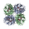
 Components Components | Neuraminidase | |||||||||
 Keywords Keywords | VIRAL PROTEIN / neuraminidase / influenza / complex / inhibitor | |||||||||
| Function / homology |  Function and homology information Function and homology informationexo-alpha-sialidase / exo-alpha-sialidase activity / viral budding from plasma membrane / carbohydrate metabolic process / host cell plasma membrane / virion membrane / membrane / metal ion binding Similarity search - Function | |||||||||
| Biological species |   unidentified influenza virus unidentified influenza virus | |||||||||
| Method |  X-RAY DIFFRACTION / MOLECULAR REPLACEMENT / Resolution: 1.69 Å X-RAY DIFFRACTION / MOLECULAR REPLACEMENT / Resolution: 1.69 Å | |||||||||
 Authors Authors | Pachl, P. / Pokorna, J. / Hejdanek, J. | |||||||||
 Citation Citation | Journal: Viruses / Year: 2018 Title: Kinetic, Thermodynamic, and Structural Analysis of Drug Resistance Mutations in Neuraminidase from the 2009 Pandemic Influenza Virus. Authors: Pokorna, J. / Pachl, P. / Karlukova, E. / Hejdanek, J. / Rezacova, P. / Machara, A. / Hudlicky, J. / Konvalinka, J. / Kozisek, M. | |||||||||
| History |
|
- Structure visualization
| Structure viewer | Molecule: MolmilJmol/JSmol |
|---|
- Downloads & links
-Download
| PDBx/mmCIF format | 5nze.cif.gz | 190.2 KB | Display | PDBx/mmCIF format |
|---|---|---|---|---|
| PDB format | pdb5nze.ent.gz | 146.4 KB | Display | PDB format |
| PDBx/mmJSON format | 5nze.json.gz | Tree view | PDBx/mmJSON format | |
| Others |  Other downloads Other downloads |
-Validation report
| Arichive directory | https://data.pdbj.org/pub/pdb/validation_reports/nz/5nzeftp://data.pdbj.org/pub/pdb/validation_reports/nz/5nze | HTTPS FTP |
|---|
-Related structure data
| Related structure data | 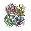 5nweC 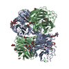 5nz4C  5nzfC  5nznC 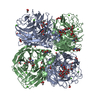 3ti6S C: citing same article ( S: Starting model for refinement |
|---|---|
| Similar structure data |


-Links
 PDBj
PDBj

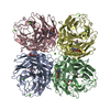
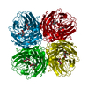
- Assembly
| Deposited unit | 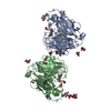
| |||||||||
|---|---|---|---|---|---|---|---|---|---|---|
| 1 | 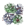
| |||||||||
| 2 | 
| |||||||||
| Unit cell |
| |||||||||
| Components on special symmetry positions |
|
-Components
-Protein , 1 types, 2 molecules AB
| #1: Protein | Mass: 42754.652 Da / Num. of mol.: 2 Source method: isolated from a genetically manipulated source Source: (gene. exp.) unidentified influenza virus / Gene: NA / Production host:  |
|---|
-Sugars , 2 types, 5 molecules 
| #2: Polysaccharide | alpha-D-mannopyranose-(1-3)-beta-D-mannopyranose-(1-4)-2-acetamido-2-deoxy-beta-D-glucopyranose-(1- ...alpha-D-mannopyranose-(1-3)-beta-D-mannopyranose-(1-4)-2-acetamido-2-deoxy-beta-D-glucopyranose-(1-4)-2-acetamido-2-deoxy-beta-D-glucopyranose Source method: isolated from a genetically manipulated source |
|---|---|
| #5: Sugar | ChemComp-NAG /  Type: D-saccharide, beta linking / Mass: 221.208 Da / Num. of mol.: 4 Type: D-saccharide, beta linking / Mass: 221.208 Da / Num. of mol.: 4Source method: isolated from a genetically manipulated source Formula: C8H15NO6 |
-Non-polymers , 4 types, 811 molecules 
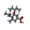


| #3: Chemical | ChemComp-CA /  Mass: 40.078 Da / Num. of mol.: 5 / Source method: obtained synthetically / Formula: Ca Mass: 40.078 Da / Num. of mol.: 5 / Source method: obtained synthetically / Formula: Ca#4: Chemical |  Mass: 284.351 Da / Num. of mol.: 2 / Source method: obtained synthetically / Formula: C14H24N2O4 / Comment: medication*YM Mass: 284.351 Da / Num. of mol.: 2 / Source method: obtained synthetically / Formula: C14H24N2O4 / Comment: medication*YM#6: Chemical | ChemComp-EDO /  Mass: 62.068 Da / Num. of mol.: 14 Mass: 62.068 Da / Num. of mol.: 14Source method: isolated from a genetically manipulated source Formula: C2H6O2 #7: Water | ChemComp-HOH / | Mass: 18.015 Da / Num. of mol.: 790 / Source method: isolated from a natural source / Formula: H2O |
|---|
-Details
| Has protein modification | Y |
|---|
-Experimental details
-Experiment
| Experiment | Method: X-RAY DIFFRACTION / Number of used crystals: 1 |
|---|
- Sample preparation
| Crystal | Density Matthews: 2.82 Å3/Da / Density % sol: 56.43 % |
|---|---|
| Crystal grow | Temperature: 291 K / Method: vapor diffusion, hanging drop / pH: 8 / Details: 0.1 M HEPES pH 6.7, 8.5% PEG 8000 |
-Data collection
| Diffraction | Mean temperature: 100 K |
|---|---|
| Diffraction source | Source: ROTATING ANODE / Type: RIGAKU MICROMAX-007 HF / Wavelength: 1.54 Å |
| Detector | Type: DECTRIS PILATUS 300K / Detector: PIXEL / Date: Sep 30, 2016 |
| Radiation | Protocol: SINGLE WAVELENGTH / Monochromatic (M) / Laue (L): M / Scattering type: x-ray |
| Radiation wavelength | Wavelength: 1.54 Å / Relative weight: 1 |
| Reflection | Resolution: 1.69→49.6 Å / Num. obs: 101435 / % possible obs: 93.9 % / Observed criterion σ(I): -3 / Redundancy: 4.8 % / Biso Wilson estimate: 22.396 Å2 / CC1/2: 0.996 / Rmerge(I) obs: 0.089 / Rrim(I) all: 0.094 / Net I/σ(I): 11.6 |
| Reflection shell | Resolution: 1.69→1.73 Å / Redundancy: 3.61 % / Rmerge(I) obs: 0.712 / Mean I/σ(I) obs: 2 / Num. unique obs: 6625 / CC1/2: 0.821 / Rrim(I) all: 0.7 / % possible all: 83.2 |
- Processing
| Software |
| ||||||||||||||||||||||||||||||||||||||||||||||||||||||||||||
|---|---|---|---|---|---|---|---|---|---|---|---|---|---|---|---|---|---|---|---|---|---|---|---|---|---|---|---|---|---|---|---|---|---|---|---|---|---|---|---|---|---|---|---|---|---|---|---|---|---|---|---|---|---|---|---|---|---|---|---|---|---|
| Refinement | Method to determine structure: MOLECULAR REPLACEMENT Starting model: 3TI6 Resolution: 1.69→49.6 Å / Cor.coef. Fo:Fc: 0.953 / Cor.coef. Fo:Fc free: 0.941 / SU B: 2.175 / SU ML: 0.07 / SU R Cruickshank DPI: 0.1021 / Cross valid method: THROUGHOUT / σ(F): 0 / ESU R: 0.102 / ESU R Free: 0.099 Details: HYDROGENS HAVE BEEN ADDED IN THE RIDING POSITIONS U VALUES : REFINED INDIVIDUALLY
| ||||||||||||||||||||||||||||||||||||||||||||||||||||||||||||
| Solvent computation | Ion probe radii: 0.8 Å / Shrinkage radii: 0.8 Å / VDW probe radii: 1.2 Å | ||||||||||||||||||||||||||||||||||||||||||||||||||||||||||||
| Displacement parameters | Biso max: 53.94 Å2 / Biso mean: 16.962 Å2 / Biso min: 5.65 Å2
| ||||||||||||||||||||||||||||||||||||||||||||||||||||||||||||
| Refinement step | Cycle: final / Resolution: 1.69→49.6 Å
| ||||||||||||||||||||||||||||||||||||||||||||||||||||||||||||
| Refine LS restraints |
| ||||||||||||||||||||||||||||||||||||||||||||||||||||||||||||
| LS refinement shell | Resolution: 1.69→1.734 Å / Rfactor Rfree error: 0 / Total num. of bins used: 20
|
