 Movie
Movie Controller

[English] 日本語
 Yorodumi
- PDB-6h9w: Unraveling the role of the secretor antigen in human rotavirus at... -
+ Open data

- Basic information
| Entry | Database: PDB / ID: 6h9w | ||||||
|---|---|---|---|---|---|---|---|
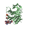
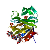
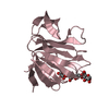
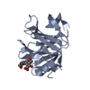
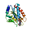
| Title | Unraveling the role of the secretor antigen in human rotavirus attachment to histo-blood group antigens | ||||||
 Components Components | Outer capsid protein VP4 | ||||||
 Keywords Keywords | VIRAL PROTEIN / histo-blood group antigen rotavirus | ||||||
| Function / homology |  Function and homology information Function and homology informationviral outer capsid / symbiont entry into host cell via permeabilization of host membrane / virion attachment to host cell Similarity search - Function | ||||||
| Biological species |  Rotavirus A Rotavirus A | ||||||
| Method |  X-RAY DIFFRACTION / SYNCHROTRON / MOLECULAR REPLACEMENT / Resolution: 1.35 Å X-RAY DIFFRACTION / SYNCHROTRON / MOLECULAR REPLACEMENT / Resolution: 1.35 Å | ||||||
 Authors Authors | Ciges-Tomas, J.R. / Gozalbo-Rovira, R. / Vila-Vicent, S. / Buesa, J. / Santiso-Bellon, C. / Monedero, V. / Yebra, M.J. / Rodriguez-Diaz, J. / Marina, A. | ||||||
| Funding support |  Spain, 1items Spain, 1items
| ||||||
 Citation Citation | Journal: Plos Pathog. / Year: 2019 Title: Unraveling the role of the secretor antigen in human rotavirus attachment to histo-blood group antigens. Authors: Gozalbo-Rovira, R. / Ciges-Tomas, J.R. / Vila-Vicent, S. / Buesa, J. / Santiso-Bellon, C. / Monedero, V. / Yebra, M.J. / Marina, A. / Rodriguez-Diaz, J. | ||||||
| History |
|
- Structure visualization
| Structure viewer | Molecule: MolmilJmol/JSmol |
|---|
- Downloads & links
-Download
| PDBx/mmCIF format | 6h9w.cif.gz | 53.9 KB | Display | PDBx/mmCIF format |
|---|---|---|---|---|
| PDB format | pdb6h9w.ent.gz | 37.4 KB | Display | PDB format |
| PDBx/mmJSON format | 6h9w.json.gz | Tree view | PDBx/mmJSON format | |
| Others |  Other downloads Other downloads |
-Validation report
| Arichive directory | https://data.pdbj.org/pub/pdb/validation_reports/h9/6h9wftp://data.pdbj.org/pub/pdb/validation_reports/h9/6h9w | HTTPS FTP |
|---|
-Related structure data
| Related structure data |  6h9yC  6h9zC  6ha0C 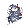 2i2sS S: Starting model for refinement C: citing same article ( |
|---|---|
| Similar structure data |

-Links
 PDBj
PDBj
- Assembly
| Deposited unit | 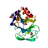
| ||||||||
|---|---|---|---|---|---|---|---|---|---|
| 1 |
| ||||||||
| Unit cell |
| ||||||||
| Components on special symmetry positions |
|
-Components
| #1: Protein | Mass: 18550.332 Da / Num. of mol.: 1 Source method: isolated from a genetically manipulated source Details: G1 and S2 are residuals from tag digestion / Source: (gene. exp.) Rotavirus A / Gene: VP4 / Production host:  | ||||||
|---|---|---|---|---|---|---|---|
| #2: Chemical | ChemComp-SO4 /   Mass: 96.063 Da / Num. of mol.: 4 / Source method: obtained synthetically / Formula: SO4 Mass: 96.063 Da / Num. of mol.: 4 / Source method: obtained synthetically / Formula: SO4#3: Chemical | ChemComp-CIT / |   Mass: 192.124 Da / Num. of mol.: 1 / Source method: obtained synthetically / Formula: C6H8O7 Mass: 192.124 Da / Num. of mol.: 1 / Source method: obtained synthetically / Formula: C6H8O7#4: Chemical |   Mass: 60.095 Da / Num. of mol.: 2 / Source method: obtained synthetically / Formula: C3H8O Mass: 60.095 Da / Num. of mol.: 2 / Source method: obtained synthetically / Formula: C3H8O#5: Water | ChemComp-HOH / |  Mass: 18.015 Da / Num. of mol.: 211 / Source method: isolated from a natural source / Formula: H2O Mass: 18.015 Da / Num. of mol.: 211 / Source method: isolated from a natural source / Formula: H2O |
-Experimental details
-Experiment
| Experiment | Method: X-RAY DIFFRACTION / Number of used crystals: 1 |
|---|
- Sample preparation
| Crystal | Density Matthews: 3.21 Å3/Da / Density % sol: 61.68 % |
|---|---|
| Crystal grow | Temperature: 294.15 K / Method: vapor diffusion, sitting drop Details: 1.2M (NH 4 ) 2 SO 4 3% iso-propanol and 0.1M sodium citrate pH4.6 |
-Data collection
| Diffraction | Mean temperature: 100 K |
|---|---|
| Diffraction source | Source: SYNCHROTRON / Site: ALBA / Beamline: XALOC / Wavelength: 0.97923 Å |
| Detector | Type: DECTRIS PILATUS 6M / Detector: PIXEL / Date: Jul 7, 2017 |
| Radiation | Protocol: SINGLE WAVELENGTH / Monochromatic (M) / Laue (L): M / Scattering type: x-ray |
| Radiation wavelength | Wavelength: 0.97923 Å / Relative weight: 1 |
| Reflection | Resolution: 1.35→65.95 Å / Num. obs: 52302 / % possible obs: 99.9 % / Redundancy: 10 % / CC1/2: 0.999 / Rpim(I) all: 0.021 / Net I/σ(I): 15.9 |
| Reflection shell | Resolution: 1.35→1.42 Å / Redundancy: 5.8 % / Mean I/σ(I) obs: 1.9 / Num. unique obs: 7527 / CC1/2: 0.932 / Rpim(I) all: 0.266 / % possible all: 99.6 |
- Processing
| Software |
| ||||||||||||||||||||||||||||||||||||||||||||||||||||||||||||
|---|---|---|---|---|---|---|---|---|---|---|---|---|---|---|---|---|---|---|---|---|---|---|---|---|---|---|---|---|---|---|---|---|---|---|---|---|---|---|---|---|---|---|---|---|---|---|---|---|---|---|---|---|---|---|---|---|---|---|---|---|---|
| Refinement | Method to determine structure: MOLECULAR REPLACEMENT Starting model: 2I2S Resolution: 1.35→65.95 Å / Cor.coef. Fo:Fc: 0.967 / Cor.coef. Fo:Fc free: 0.961 / SU B: 1.201 / SU ML: 0.048 / Cross valid method: THROUGHOUT / σ(F): 0 / ESU R: 0.054 / ESU R Free: 0.055 Details: HYDROGENS HAVE BEEN ADDED IN THE RIDING POSITIONS U VALUES : REFINED INDIVIDUALLY
| ||||||||||||||||||||||||||||||||||||||||||||||||||||||||||||
| Solvent computation | Ion probe radii: 0.8 Å / Shrinkage radii: 0.8 Å / VDW probe radii: 1.2 Å | ||||||||||||||||||||||||||||||||||||||||||||||||||||||||||||
| Displacement parameters | Biso max: 79.2 Å2 / Biso mean: 27.175 Å2 / Biso min: 14.52 Å2
| ||||||||||||||||||||||||||||||||||||||||||||||||||||||||||||
| Refinement step | Cycle: final / Resolution: 1.35→65.95 Å
| ||||||||||||||||||||||||||||||||||||||||||||||||||||||||||||
| Refine LS restraints |
| ||||||||||||||||||||||||||||||||||||||||||||||||||||||||||||
| LS refinement shell | Resolution: 1.347→1.382 Å / Rfactor Rfree error: 0 / Total num. of bins used: 20
|
