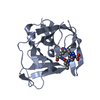
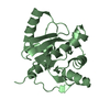
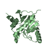
Entry Database : PDB / ID : 1b1cTitle CRYSTAL STRUCTURE OF THE FMN-BINDING DOMAIN OF HUMAN CYTOCHROME P450 REDUCTASE AT 1.93A RESOLUTION PROTEIN (NADPH-CYTOCHROME P450 REDUCTASE) Keywords / / / / / Function / homology Function Domain/homology Component
/ / / / / / / / / / / / / / / / / / / / / / / / / / / / / / / / / / / / / / / / / / / / / / / / / / / / / / / / / / / / / / / / / / / / / / / / / / Biological species Homo sapiens (human)Method / / Resolution : 1.93 Å Authors Zhao, Q. / Modi, S. / Smith, G. / Paine, M. / Mcdonagh, P.D. / Wolf, C.R. / Tew, D. / Lian, L.-Y. / Roberts, G.C.K. / Driessen, H.P.C. Journal : Protein Sci. / Year : 1999Title : Crystal structure of the FMN-binding domain of human cytochrome P450 reductase at 1.93 A resolution.
Authors :
Zhao, Q. / Modi, S. / Smith, G. / Paine, M. / McDonagh, P.D. / Wolf, C.R. / Tew, D. / Lian, L.Y. / Roberts, G.C. / Driessen, H.P. #1: Journal : J.Struct.Biol. / Year : 1996Title : Crystallization and Preliminary X-Ray Diffraction Studies of Human Cytochrome P450 Reductase
Authors :
Zhao, Q. / Smith, G. / Modi, S. / Paine, M. / Wolf, R.C. / Tew, D. / Lian, L.Y. / Primrose, W.U. / Roberts, G.C. / Driessen, H.P. History Deposition Nov 19, 1998 Deposition site / Processing site Revision 1.0 Nov 24, 1999 Provider / Type Revision 1.1 Apr 26, 2008 Group Revision 1.2 Jul 13, 2011 Group Revision 1.3 Oct 4, 2017 Group / Category / Item Revision 1.4 Dec 27, 2023 Group / Database references / Derived calculationsCategory chem_comp_atom / chem_comp_bond ... chem_comp_atom / chem_comp_bond / database_2 / pdbx_struct_conn_angle / struct_conn / struct_site Item _database_2.pdbx_DOI / _database_2.pdbx_database_accession ... _database_2.pdbx_DOI / _database_2.pdbx_database_accession / _pdbx_struct_conn_angle.ptnr1_auth_comp_id / _pdbx_struct_conn_angle.ptnr1_auth_seq_id / _pdbx_struct_conn_angle.ptnr1_label_asym_id / _pdbx_struct_conn_angle.ptnr1_label_atom_id / _pdbx_struct_conn_angle.ptnr1_label_comp_id / _pdbx_struct_conn_angle.ptnr1_label_seq_id / _pdbx_struct_conn_angle.ptnr1_symmetry / _pdbx_struct_conn_angle.ptnr3_auth_comp_id / _pdbx_struct_conn_angle.ptnr3_auth_seq_id / _pdbx_struct_conn_angle.ptnr3_label_asym_id / _pdbx_struct_conn_angle.ptnr3_label_atom_id / _pdbx_struct_conn_angle.ptnr3_label_comp_id / _pdbx_struct_conn_angle.ptnr3_label_seq_id / _pdbx_struct_conn_angle.ptnr3_symmetry / _pdbx_struct_conn_angle.value / _struct_conn.pdbx_dist_value / _struct_conn.ptnr1_auth_comp_id / _struct_conn.ptnr1_auth_seq_id / _struct_conn.ptnr1_label_asym_id / _struct_conn.ptnr1_label_atom_id / _struct_conn.ptnr1_label_comp_id / _struct_conn.ptnr1_label_seq_id / _struct_conn.ptnr1_symmetry / _struct_conn.ptnr2_auth_comp_id / _struct_conn.ptnr2_auth_seq_id / _struct_conn.ptnr2_label_asym_id / _struct_conn.ptnr2_label_atom_id / _struct_conn.ptnr2_label_comp_id / _struct_conn.ptnr2_label_seq_id / _struct_conn.ptnr2_symmetry / _struct_site.pdbx_auth_asym_id / _struct_site.pdbx_auth_comp_id / _struct_site.pdbx_auth_seq_id
Show all Show less
 Movie
Movie Controller

 Yorodumi
 Open data
 Basic information
 Components
 Keywords
 Function and homology information
 Homo sapiens (human)
 X-RAY DIFFRACTION /
  Authors
 Citation
 Structure visualization
 Downloads & links
 Other downloads
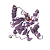


 PDBj
PDBj

 Assembly
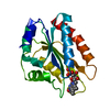

 Mass: 40.078 Da / Num. of mol.: 1 / Source method: obtained synthetically / Formula: Ca
 Mass: 456.344 Da / Num. of mol.: 1 / Source method: obtained synthetically / Formula: C17H21N4O9P
 Mass: 18.015 Da / Num. of mol.: 61 / Source method: isolated from a natural source / Formula: H2O
 Sample preparation
 Processing