 Movie
Movie Controller

[English] 日本語
 Yorodumi
- PDB-6h86: Rebuilt and re-refined PDB entry 4R3Q: Crystal structure of SYCE3 -
+ Open data

- Basic information
| Entry | Database: PDB / ID: 6h86 | |||||||||
|---|---|---|---|---|---|---|---|---|---|---|
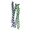
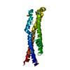
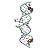
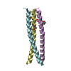
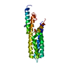
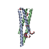
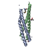
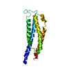
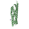
| Title | Rebuilt and re-refined PDB entry 4R3Q: Crystal structure of SYCE3 | |||||||||
 Components Components | Synaptonemal complex central element protein 3 | |||||||||
 Keywords Keywords | STRUCTURAL PROTEIN / Meiosis / synaptonemal complex | |||||||||
| Function / homology |  Function and homology information Function and homology informationcentral element / synaptonemal complex assembly / reciprocal meiotic recombination / chromosome / spermatogenesis / positive regulation of apoptotic process / cell division / nucleoplasm / nucleus Similarity search - Function | |||||||||
| Biological species |  | |||||||||
| Method |  X-RAY DIFFRACTION / SYNCHROTRON / Resolution: 1.901 Å X-RAY DIFFRACTION / SYNCHROTRON / Resolution: 1.901 Å | |||||||||
 Authors Authors | Davies, O.R. | |||||||||
| Funding support |  United Kingdom, 2items United Kingdom, 2items
| |||||||||
 Citation Citation | Journal: J.Biol.Chem. / Year: 2019 Title: A molecular model for self-assembly of the synaptonemal complex protein SYCE3. Authors: Dunne, O.M. / Davies, O.R. | |||||||||
| History |
|
- Structure visualization
| Structure viewer | Molecule: MolmilJmol/JSmol |
|---|
- Downloads & links
-Download
| PDBx/mmCIF format | 6h86.cif.gz | 108.5 KB | Display | PDBx/mmCIF format |
|---|---|---|---|---|
| PDB format | pdb6h86.ent.gz | 86.9 KB | Display | PDB format |
| PDBx/mmJSON format | 6h86.json.gz | Tree view | PDBx/mmJSON format | |
| Others |  Other downloads Other downloads |
-Validation report
| Arichive directory | https://data.pdbj.org/pub/pdb/validation_reports/h8/6h86ftp://data.pdbj.org/pub/pdb/validation_reports/h8/6h86 | HTTPS FTP |
|---|
-Related structure data
| Related structure data | |
|---|---|
| Similar structure data |


-Links
 PDBj
PDBj- Assembly
| Deposited unit | 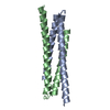
| ||||||||
|---|---|---|---|---|---|---|---|---|---|
| 1 |
| ||||||||
| Unit cell |
| ||||||||
| Components on special symmetry positions |
|
-Components
| #1: Protein | Mass: 10480.013 Da / Num. of mol.: 2 Source method: isolated from a genetically manipulated source Source: (gene. exp.)  #2: Water | ChemComp-HOH / |  Mass: 18.015 Da / Num. of mol.: 73 / Source method: isolated from a natural source / Formula: H2O Mass: 18.015 Da / Num. of mol.: 73 / Source method: isolated from a natural source / Formula: H2O |
|---|
-Experimental details
-Experiment
| Experiment | Method: X-RAY DIFFRACTION / Number of used crystals: 1 |
|---|
- Sample preparation
| Crystal | Density Matthews: 2.66 Å3/Da / Density % sol: 53.79 % |
|---|---|
| Crystal grow | Temperature: 277 K / Method: vapor diffusion, sitting drop Details: 0.1M Citric acid, 7% 2-propanol, 1% PEG 20000, pH 3.5, VAPOR DIFFUSION, SITTING DROP, temperature 277K |
-Data collection
| Diffraction | Mean temperature: 100 K |
|---|---|
| Diffraction source | Source: SYNCHROTRON / Site: SSRF  / Beamline: BL17U / Wavelength: 0.9796 Å / Beamline: BL17U / Wavelength: 0.9796 Å |
| Detector | Type: MARMOSAIC 225 mm CCD / Detector: CCD / Date: Jun 30, 2013 |
| Radiation | Protocol: SINGLE WAVELENGTH / Monochromatic (M) / Laue (L): M / Scattering type: x-ray |
| Radiation wavelength | Wavelength: 0.9796 Å / Relative weight: 1 |
| Reflection | Resolution: 1.9→50 Å / Num. obs: 16498 / % possible obs: 96.89 % / Redundancy: 3.9 % / Rsym value: 0.059 / Net I/σ(I): 22 |
| Reflection shell | Resolution: 1.9→1.93 Å / Rsym value: 0.486 |
- Processing
| Software |
| |||||||||||||||||||||||||||||||||||||||||||||||||
|---|---|---|---|---|---|---|---|---|---|---|---|---|---|---|---|---|---|---|---|---|---|---|---|---|---|---|---|---|---|---|---|---|---|---|---|---|---|---|---|---|---|---|---|---|---|---|---|---|---|---|
| Refinement | Resolution: 1.901→23.661 Å / SU ML: 0.2 / Cross valid method: FREE R-VALUE / σ(F): 2.01 / Phase error: 25.32
| |||||||||||||||||||||||||||||||||||||||||||||||||
| Solvent computation | Shrinkage radii: 0.9 Å / VDW probe radii: 1.11 Å | |||||||||||||||||||||||||||||||||||||||||||||||||
| Refinement step | Cycle: LAST / Resolution: 1.901→23.661 Å
| |||||||||||||||||||||||||||||||||||||||||||||||||
| Refine LS restraints |
| |||||||||||||||||||||||||||||||||||||||||||||||||
| LS refinement shell |
| |||||||||||||||||||||||||||||||||||||||||||||||||
| Refinement TLS params. | Method: refined / Origin x: 31.5493 Å / Origin y: -11.0764 Å / Origin z: -0.2192 Å
| |||||||||||||||||||||||||||||||||||||||||||||||||
| Refinement TLS group | Selection details: all |
