 Movie
Movie Controller


+ Open data

- Basic information
| Entry | Database: PDB / ID: 1vls | ||||||
|---|---|---|---|---|---|---|---|
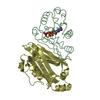
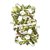
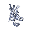
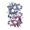
| Title | LIGAND BINDING DOMAIN OF THE WILD-TYPE ASPARTATE RECEPTOR | ||||||
 Components Components | ASPARTATE RECEPTOR | ||||||
 Keywords Keywords | CHEMOTAXIS / BACTERIAL CHEMOTAXIS RECEPTOR / UNBOUND | ||||||
| Function / homology |  Function and homology information Function and homology informationchemotaxis / transmembrane signaling receptor activity / signal transduction / plasma membrane Similarity search - Function | ||||||
| Biological species |  Salmonella typhimurium (bacteria) Salmonella typhimurium (bacteria) | ||||||
| Method |  X-RAY DIFFRACTION / Resolution: 1.85 Å X-RAY DIFFRACTION / Resolution: 1.85 Å | ||||||
 Authors Authors | Kim, S.-H. / Yeh, J.I. / Biemann, H.-P. / Prive, G. / Pandit, J. / Koshland Junior, D.E. | ||||||
 Citation Citation | Journal: J.Mol.Biol. / Year: 1996 Title: High-resolution structures of the ligand binding domain of the wild-type bacterial aspartate receptor. Authors: Yeh, J.I. / Biemann, H.P. / Prive, G.G. / Pandit, J. / Koshland Jr., D.E. / Kim, S.H. #1: Journal: J.Biol.Chem. / Year: 1993Title: The Three-Dimensional Structure of the Ligand-Binding Domain of a Wild-Type Bacterial Chemotaxis Receptor. Structural Comparison to the Cross-Linked Mutant Forms and Conformational Changes Upon Ligand Binding Authors: Yeh, J.I. / Biemann, H.P. / Pandit, J. / Koshland, D.E. / Kim, S.H. #2: Journal: Science / Year: 1991Title: Three-Dimensional Structures of the Ligand-Binding Domain of the Bacterial Aspartate Receptor with and without a Ligand Authors: Milburn, M.V. / Prive, G.G. / Milligan, D.L. / Scott, W.G. / Yeh, J. / Jancarik, J. / Koshland Junior, D.E. / Kim, S.H. | ||||||
| History |
|
- Structure visualization
| Structure viewer | Molecule: MolmilJmol/JSmol |
|---|
- Downloads & links
-Download
| PDBx/mmCIF format | 1vls.cif.gz | 44.8 KB | Display | PDBx/mmCIF format |
|---|---|---|---|---|
| PDB format | pdb1vls.ent.gz | 31.9 KB | Display | PDB format |
| PDBx/mmJSON format | 1vls.json.gz | Tree view | PDBx/mmJSON format | |
| Others |  Other downloads Other downloads |
-Validation report
| Arichive directory | https://data.pdbj.org/pub/pdb/validation_reports/vl/1vlsftp://data.pdbj.org/pub/pdb/validation_reports/vl/1vls | HTTPS FTP |
|---|
-Related structure data


-Links
 PDBj
PDBj

- Assembly
| Deposited unit | 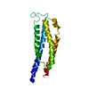
| ||||||||
|---|---|---|---|---|---|---|---|---|---|
| 1 | 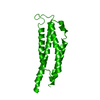
| ||||||||
| Unit cell |
|
-Components
| #1: Protein | Mass: 16366.229 Da / Num. of mol.: 1 Source method: isolated from a genetically manipulated source Source: (gene. exp.) Salmonella typhimurium (bacteria) / References: UniProt: P02941 |
|---|---|
| #2: Water | ChemComp-HOH /  Mass: 18.015 Da / Num. of mol.: 219 / Source method: isolated from a natural source / Formula: H2O Mass: 18.015 Da / Num. of mol.: 219 / Source method: isolated from a natural source / Formula: H2O |
-Experimental details
-Experiment
| Experiment | Method: X-RAY DIFFRACTION |
|---|
- Sample preparation
| Crystal | Density Matthews: 2.34 Å3/Da / Density % sol: 42 % | ||||||||||||||||||||||||||||||||||||||||||||||||
|---|---|---|---|---|---|---|---|---|---|---|---|---|---|---|---|---|---|---|---|---|---|---|---|---|---|---|---|---|---|---|---|---|---|---|---|---|---|---|---|---|---|---|---|---|---|---|---|---|---|
| Crystal grow | *PLUS Method: vapor diffusion | ||||||||||||||||||||||||||||||||||||||||||||||||
| Components of the solutions | *PLUS
|
-Data collection
| Diffraction source | Wavelength: 1.5418 |
|---|---|
| Detector | Type: RIGAKU / Detector: IMAGE PLATE |
| Radiation | Monochromatic (M) / Laue (L): M / Scattering type: x-ray |
| Radiation wavelength | Wavelength: 1.5418 Å / Relative weight: 1 |
| Reflection | Redundancy: 4.2 % / Rmerge(I) obs: 0.072 |
| Reflection | *PLUS Highest resolution: 1.85 Å / Lowest resolution: 6 Å / Num. obs: 12189 / Num. measured all: 51445 |
- Processing
| Software |
| ||||||||||||
|---|---|---|---|---|---|---|---|---|---|---|---|---|---|
| Refinement | Resolution: 1.85→6 Å / σ(F): 1
| ||||||||||||
| Displacement parameters | Biso mean: 32.7 Å2 | ||||||||||||
| Refinement step | Cycle: LAST / Resolution: 1.85→6 Å
| ||||||||||||
| Software | *PLUS Name: X-PLOR / Classification: refinement | ||||||||||||
| Refine LS restraints | *PLUS
|
