 Movie
Movie Controller


+ Open data

- Basic information
| Entry | Database: PDB / ID: 6h7w | ||||||||||||||||||||||||
|---|---|---|---|---|---|---|---|---|---|---|---|---|---|---|---|---|---|---|---|---|---|---|---|---|---|
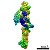
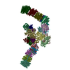
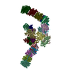
| Title | Model of retromer-Vps5 complex assembled on membrane. | ||||||||||||||||||||||||
 Components Components |
| ||||||||||||||||||||||||
 Keywords Keywords | PROTEIN TRANSPORT / retromer / endosome / sorting nexin / BAR / membrane trafficking | ||||||||||||||||||||||||
| Function / homology |  Function and homology information Function and homology informationretromer, cargo-selective complex / protein retention in Golgi apparatus / retromer complex / retrograde transport, endosome to Golgi / phosphatidylinositol binding / intracellular protein transport / late endosome / protein transport / endosome / Golgi apparatus ...retromer, cargo-selective complex / protein retention in Golgi apparatus / retromer complex / retrograde transport, endosome to Golgi / phosphatidylinositol binding / intracellular protein transport / late endosome / protein transport / endosome / Golgi apparatus / identical protein binding / cytosol Similarity search - Function | ||||||||||||||||||||||||
| Biological species |  Chaetomium thermophilum (fungus) Chaetomium thermophilum (fungus) | ||||||||||||||||||||||||
| Method | ELECTRON MICROSCOPY / subtomogram averaging / cryo EM / Resolution: 11.4 Å | ||||||||||||||||||||||||
 Authors Authors | Kovtun, O. / Leneva, N. / Ariotti, N. / Rohan, T.S. / Owen, D.J. / Briggs, J.A.G. / Collins, B.M. | ||||||||||||||||||||||||
| Funding support |  United Kingdom, United Kingdom,  Australia, 7items Australia, 7items
| ||||||||||||||||||||||||
 Citation Citation | Journal: Nature / Year: 2018 Title: Structure of the membrane-assembled retromer coat determined by cryo-electron tomography. Authors: Oleksiy Kovtun / Natalya Leneva / Yury S Bykov / Nicholas Ariotti / Rohan D Teasdale / Miroslava Schaffer / Benjamin D Engel / David J Owen / John A G Briggs / Brett M Collins /  Abstract: Eukaryotic cells traffic proteins and lipids between different compartments using protein-coated vesicles and tubules. The retromer complex is required to generate cargo-selective tubulovesicular ...Eukaryotic cells traffic proteins and lipids between different compartments using protein-coated vesicles and tubules. The retromer complex is required to generate cargo-selective tubulovesicular carriers from endosomal membranes. Conserved in eukaryotes, retromer controls the cellular localization and homeostasis of hundreds of transmembrane proteins, and its disruption is associated with major neurodegenerative disorders. How retromer is assembled and how it is recruited to form coated tubules is not known. Here we describe the structure of the retromer complex (Vps26-Vps29-Vps35) assembled on membrane tubules with the bin/amphiphysin/rvs-domain-containing sorting nexin protein Vps5, using cryo-electron tomography and subtomogram averaging. This reveals a membrane-associated Vps5 array, from which arches of retromer extend away from the membrane surface. Vps35 forms the 'legs' of these arches, and Vps29 resides at the apex where it is free to interact with regulatory factors. The bases of the arches connect to each other and to Vps5 through Vps26, and the presence of the same arches on coated tubules within cells confirms their functional importance. Vps5 binds to Vps26 at a position analogous to the previously described cargo- and Snx3-binding site, which suggests the existence of distinct retromer-sorting nexin assemblies. The structure provides insight into the architecture of the coat and its mechanism of assembly, and suggests that retromer promotes tubule formation by directing the distribution of sorting nexin proteins on the membrane surface while providing a scaffold for regulatory-protein interactions. | ||||||||||||||||||||||||
| History |
|
- Structure visualization
| Movie |
Movie viewer |
|---|---|
| Structure viewer | Molecule: MolmilJmol/JSmol |
- Downloads & links
-Download
| PDBx/mmCIF format | 6h7w.cif.gz | 1.9 MB | Display | PDBx/mmCIF format |
|---|---|---|---|---|
| PDB format | pdb6h7w.ent.gz | 1.6 MB | Display | PDB format |
| PDBx/mmJSON format | 6h7w.json.gz | Tree view | PDBx/mmJSON format | |
| Others |  Other downloads Other downloads |
-Validation report
| Arichive directory | https://data.pdbj.org/pub/pdb/validation_reports/h7/6h7wftp://data.pdbj.org/pub/pdb/validation_reports/h7/6h7w | HTTPS FTP |
|---|
-Related structure data
| Related structure data | 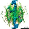 0154MC 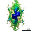 0155C 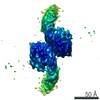 0156C 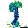 0157C 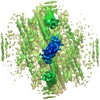 0158C 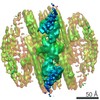 0159C 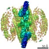 0160C 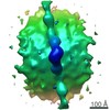 0161C 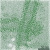 0162C 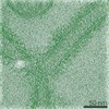 0163C 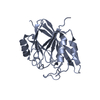 5w8mC M: map data used to model this data C: citing same article ( |
|---|---|
| Similar structure data |


-Links
 PDBj
PDBj

- Assembly
| Deposited unit | 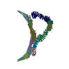
|
|---|---|
| 1 |
|
-Components
-Vacuolar protein sorting-associated protein ... , 3 types, 6 molecules JCSTQR
| #1: Protein | Mass: 34308.449 Da / Num. of mol.: 2 Source method: isolated from a genetically manipulated source Source: (gene. exp.) Chaetomium thermophilum (strain DSM 1495 / CBS 144.50 / IMI 039719) (fungus)Strain: DSM 1495 / CBS 144.50 / IMI 039719 / Gene: CTHT_0009750 / Production host:  #5: Protein | Mass: 21470.654 Da / Num. of mol.: 2 Source method: isolated from a genetically manipulated source Source: (gene. exp.) Chaetomium thermophilum (strain DSM 1495 / CBS 144.50 / IMI 039719) (fungus)Strain: DSM 1495 / CBS 144.50 / IMI 039719 / Gene: CTHT_0002370 / Production host: #6: Protein | Mass: 96140.016 Da / Num. of mol.: 2 Source method: isolated from a genetically manipulated source Source: (gene. exp.) Chaetomium thermophilum (strain DSM 1495 / CBS 144.50 / IMI 039719) (fungus)Strain: DSM 1495 / CBS 144.50 / IMI 039719 / Gene: CTHT_0035730 / Production host: |
|---|
-Putative vacuolar protein sorting-associated ... , 3 types, 14 molecules BAGEPNHFDKVLOM
| #2: Protein | Mass: 42060.762 Da / Num. of mol.: 8 Source method: isolated from a genetically manipulated source Source: (gene. exp.) Chaetomium thermophilum (strain DSM 1495 / CBS 144.50 / IMI 039719) (fungus)Strain: DSM 1495 / CBS 144.50 / IMI 039719 / Gene: CTHT_0068290 / Production host: #3: Protein | Mass: 14897.982 Da / Num. of mol.: 4 Source method: isolated from a genetically manipulated source Source: (gene. exp.) Chaetomium thermophilum (strain DSM 1495 / CBS 144.50 / IMI 039719) (fungus)Strain: DSM 1495 / CBS 144.50 / IMI 039719 / Gene: CTHT_0068290 / Production host: #4: Protein | Mass: 25477.904 Da / Num. of mol.: 2 Source method: isolated from a genetically manipulated source Source: (gene. exp.) Chaetomium thermophilum (strain DSM 1495 / CBS 144.50 / IMI 039719) (fungus)Strain: DSM 1495 / CBS 144.50 / IMI 039719 / Gene: CTHT_0068290 / Production host: |
|---|
-Experimental details
-Experiment
| Experiment | Method: ELECTRON MICROSCOPY |
|---|---|
| EM experiment | Aggregation state: 3D ARRAY / 3D reconstruction method: subtomogram averaging |
- Sample preparation
| Component | Name: Retromer-Vps5 complex assembled on the membrane. / Type: COMPLEX Details: Vps26/Vps35/Vps29 trimer recruited to the membrane via Vps5 PX-BAR protein. Entity ID: all / Source: RECOMBINANT | |||||||||||||||
|---|---|---|---|---|---|---|---|---|---|---|---|---|---|---|---|---|
| Molecular weight | Units: KILODALTONS/NANOMETER / Experimental value: NO | |||||||||||||||
| Source (natural) | Organism: Chaetomium thermophilum var. thermophilum DSM 1495 (fungus) | |||||||||||||||
| Source (recombinant) | Organism: | |||||||||||||||
| Buffer solution | pH: 7.5 | |||||||||||||||
| Buffer component |
| |||||||||||||||
| Specimen | Conc.: 1.1 mg/ml / Embedding applied: NO / Shadowing applied: NO / Staining applied: NO / Vitrification applied: YES Details: The solution-assembled complex was incubated with Folch liposomes at room temperature. | |||||||||||||||
| Vitrification | Instrument: LEICA EM GP / Cryogen name: ETHANE / Humidity: 95 % / Chamber temperature: 292 K |
- Electron microscopy imaging
| Experimental equipment |  Model: Titan Krios / Image courtesy: FEI Company |
|---|---|
| Microscopy | Model: FEI TITAN KRIOS |
| Electron gun | Electron source:  FIELD EMISSION GUN / Accelerating voltage: 300 kV / Illumination mode: FLOOD BEAM FIELD EMISSION GUN / Accelerating voltage: 300 kV / Illumination mode: FLOOD BEAM |
| Electron lens | Mode: BRIGHT FIELD / Nominal magnification: 105000 X / Nominal defocus max: 6500 nm / Nominal defocus min: 2500 nm / Cs: 2.7 mm / C2 aperture diameter: 70 µm / Alignment procedure: ZEMLIN TABLEAU |
| Specimen holder | Cryogen: NITROGEN / Specimen holder model: FEI TITAN KRIOS AUTOGRID HOLDER |
| Image recording | Electron dose: 3.17 e/Å2 / Detector mode: SUPER-RESOLUTION / Film or detector model: GATAN K2 QUANTUM (4k x 4k) / Num. of grids imaged: 2 Details: Tomographic tilt series were acquired with the dose-symmetric tilt-scheme (Hagen et al., J Struct Biol. 2017) |
| EM imaging optics | Energyfilter name: GIF Quantum LS / Energyfilter slit width: 20 eV |
| Image scans | Movie frames/image: 10 |
- Processing
| EM software |
| ||||||||||||||||||||||||||||||||
|---|---|---|---|---|---|---|---|---|---|---|---|---|---|---|---|---|---|---|---|---|---|---|---|---|---|---|---|---|---|---|---|---|---|
| Image processing | Details: After motion correction in "alignframes" (IMOD), each of the images in the tilt series was low-pass filtered according to the electron-dose acquired by the sample (Grant and Grigorieff, 2015). | ||||||||||||||||||||||||||||||||
| CTF correction | Details: CTF correction was performed with ctfphaseflip IMOD command using defocus values measured by CTFFIND4 on non-dose-filtered images. Type: PHASE FLIPPING AND AMPLITUDE CORRECTION | ||||||||||||||||||||||||||||||||
| Symmetry | Point symmetry: C2 (2 fold cyclic) | ||||||||||||||||||||||||||||||||
| 3D reconstruction | Resolution: 11.4 Å / Resolution method: FSC 0.143 CUT-OFF / Num. of particles: 16037 / Algorithm: BACK PROJECTION / Symmetry type: POINT | ||||||||||||||||||||||||||||||||
| EM volume selection | Method: geometrical seeding alogn the surface of manually traced tubules Num. of tomograms: 71 / Num. of volumes extracted: 194885 / Reference model: reference-free | ||||||||||||||||||||||||||||||||
| Atomic model building | Protocol: FLEXIBLE FIT Details: Note that the associated PDB model was not generated by fitting into this map! It was generated by fitting into the associated local high-resolution maps and then combined. Initial global ...Details: Note that the associated PDB model was not generated by fitting into this map! It was generated by fitting into the associated local high-resolution maps and then combined. Initial global ridgid body fitting was done using Chimera, followed by MDFF within NAMD for flexible fitting. Note that side chain positions are not reliable in model generated by refinement into a map at this resolution. |
