| Entry | Database: PDB / ID: 6h49
|
|---|
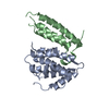
| Title | A polyamorous repressor: deciphering the evolutionary strategy used by the phage-inducible chromosomal islands to spread in nature. |
|---|
 Components Components | Orf20 |
|---|
 Keywords Keywords | STRUCTURAL PROTEIN / SaPI / Repressor |
|---|
| Function / homology | Helix-turn-helix / Helix-turn-helix XRE-family like proteins / Cro/C1-type HTH domain profile. / Cro/C1-type helix-turn-helix domain / Lambda repressor-like, DNA-binding domain superfamily / DNA binding / Orf20 Function and homology information Function and homology information |
|---|
| Biological species |   Staphylococcus aureus (bacteria) Staphylococcus aureus (bacteria) |
|---|
| Method |  X-RAY DIFFRACTION / SYNCHROTRON / MOLECULAR REPLACEMENT / Resolution: 1.8 Å X-RAY DIFFRACTION / SYNCHROTRON / MOLECULAR REPLACEMENT / Resolution: 1.8 Å |
|---|
 Authors Authors | Ciges-Tomas, J.R. / Alite, C. / Bowring, J.Z. / Donderis, J. / Penades, J.R. / Marina, A. |
|---|
| Funding support |  Spain, Spain,  United Kingdom, 6items United Kingdom, 6items | Organization | Grant number | Country |
|---|
| Spanish Ministry of Economy and Competitiveness | BIO2016-78571-P | Spain | | Spanish Ministry of Economy and Competitiveness | FPU13/02880 | Spain | | Spanish Ministry of Economy and Competitiveness | BES-2014-068617 | Spain | | Medical Research Council (United Kingdom) | MR/M003876/1 | United Kingdom | | Biotechnology and Biological Sciences Research Council | BB/N002873/1 | United Kingdom | | European Research Council | ERC-ADG/2014 670932 | United Kingdom |
|
|---|
 Citation Citation | Journal: Nat Commun / Year: 2019
Title: The structure of a polygamous repressor reveals how phage-inducible chromosomal islands spread in nature.
Authors: Rafael Ciges-Tomas, J. / Alite, C. / Humphrey, S. / Donderis, J. / Bowring, J. / Salvatella, X. / Penades, J.R. / Marina, A. |
|---|
| History | | Deposition | Jul 20, 2018 | Deposition site: PDBE / Processing site: PDBE |
|---|
| Revision 1.0 | Aug 28, 2019 | Provider: repository / Type: Initial release |
|---|
| Revision 1.1 | Oct 16, 2019 | Group: Data collection / Derived calculations / Category: reflns_shell / struct_conn / Item: _reflns_shell.d_res_high / _reflns_shell.d_res_low |
|---|
| Revision 1.2 | May 15, 2024 | Group: Data collection / Database references / Category: chem_comp_atom / chem_comp_bond / database_2
Item: _database_2.pdbx_DOI / _database_2.pdbx_database_accession |
|---|
|
|---|
 Movie
Movie Controller

 Yorodumi
 Open data
 Basic information
 Structure visualization
 Downloads & links
 Other downloads


 PDBj
PDBj


 Assembly

 Mass: 96.063 Da / Num. of mol.: 2 / Source method: obtained synthetically / Formula: SO4
 Mass: 18.015 Da / Num. of mol.: 85 / Source method: isolated from a natural source / Formula: H2O
 Sample preparation
 Processing