 Movie
Movie Controller

[English] 日本語
 Yorodumi
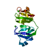
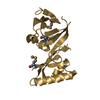
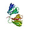
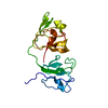
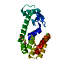
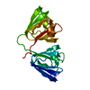
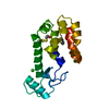
- PDB-1prr: NMR-DERIVED THREE-DIMENSIONAL SOLUTION STRUCTURE OF PROTEIN S COM... -
+ Open data

- Basic information
| Entry | Database: PDB / ID: 1prr | ||||||
|---|---|---|---|---|---|---|---|
| Title | NMR-DERIVED THREE-DIMENSIONAL SOLUTION STRUCTURE OF PROTEIN S COMPLEXED WITH CALCIUM | ||||||
 Components Components | DEVELOPMENT-SPECIFIC PROTEIN S | ||||||
 Keywords Keywords | BINDING PROTEIN | ||||||
| Function / homology |  Function and homology information Function and homology informationsporulation resulting in formation of a cellular spore / metal ion binding Similarity search - Function | ||||||
| Biological species |  Myxococcus xanthus (bacteria) Myxococcus xanthus (bacteria) | ||||||
| Method | SOLUTION NMR | ||||||
 Authors Authors | Bagby, S. / Harvey, T.S. / Eagle, S.G. / Inouye, S. / Ikura, M. | ||||||
 Citation Citation | Journal: Structure / Year: 1994 Title: NMR-derived three-dimensional solution structure of protein S complexed with calcium. Authors: Bagby, S. / Harvey, T.S. / Eagle, S.G. / Inouye, S. / Ikura, M. #1: Journal: Proc.Natl.Acad.Sci.USA / Year: 1994Title: Structural Similarity of a Developmentally Regulated Bacterial Spore Coat Protein to Betagamma-Crystallins of the Vertebrate Eye Lens Authors: Bagby, S. / Harvey, T.S. / Eagle, S.G. / Inouye, S. / Ikura, M. #2: Journal: Biochemistry / Year: 1994Title: Unusual Helix-Containing Greek Keys in Development-Specific Ca2+-Binding Protein S. 1H, 15N and 13C Assignments and Secondary Structure Determined Using Multidimensional Double and Triple ...Title: Unusual Helix-Containing Greek Keys in Development-Specific Ca2+-Binding Protein S. 1H, 15N and 13C Assignments and Secondary Structure Determined Using Multidimensional Double and Triple Resonance Heteronuclear NMR Spectroscopy Authors: Bagby, S. / Harvey, T.S. / Kay, L.E. / Eagle, S.G. / Inouye, S. / Ikura, M. | ||||||
| History |
|
- Structure visualization
| Structure viewer | Molecule: MolmilJmol/JSmol |
|---|
- Downloads & links
-Download
| PDBx/mmCIF format | 1prr.cif.gz | 64.2 KB | Display | PDBx/mmCIF format |
|---|---|---|---|---|
| PDB format | pdb1prr.ent.gz | 48.1 KB | Display | PDB format |
| PDBx/mmJSON format | 1prr.json.gz | Tree view | PDBx/mmJSON format | |
| Others |  Other downloads Other downloads |
-Validation report
| Arichive directory | https://data.pdbj.org/pub/pdb/validation_reports/pr/1prrftp://data.pdbj.org/pub/pdb/validation_reports/pr/1prr | HTTPS FTP |
|---|
-Related structure data


-Links
 PDBj
PDBj
- Assembly
| Deposited unit | 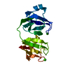
| |||||||||
|---|---|---|---|---|---|---|---|---|---|---|
| 1 |
| |||||||||
| Atom site foot note | 1: SURFACE LOOPS AT RESIDUES 33 - 40, 72 - 80, 123 - 129, AND 159 - 168 ARE RELATIVELY POORLY DEFINED. | |||||||||
| NMR ensembles |
|
-Components
| #1: Protein | Mass: 18805.865 Da / Num. of mol.: 1 Source method: isolated from a genetically manipulated source Source: (gene. exp.) Myxococcus xanthus (bacteria) / References: UniProt: P02966 |
|---|---|
| #2: Chemical |   Mass: 40.078 Da / Num. of mol.: 2 / Source method: obtained synthetically / Formula: Ca Mass: 40.078 Da / Num. of mol.: 2 / Source method: obtained synthetically / Formula: Ca |
-Experimental details
-Experiment
| Experiment | Method: SOLUTION NMR |
|---|
- Processing
| Software |
| ||||||||
|---|---|---|---|---|---|---|---|---|---|
| NMR software | Name:  X-PLOR / Developer: BRUNGER / Classification: refinement X-PLOR / Developer: BRUNGER / Classification: refinement | ||||||||
| NMR ensemble | Conformers submitted total number: 1 |
