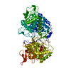
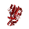
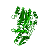
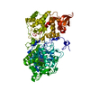
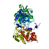
Entry Database : PDB / ID : 6h0pTitle The structure of C100A mutant of Arabidopsis thaliana UDP-apiose/UDP-xylose synthase in complex with NADH and UDP-D-glucuronic acid UDP-D-apiose/UDP-D-xylose synthase 1 Keywords / / / / / Function / homology Function Domain/homology Component
/ / / / / / / / / / / / / / / / / / / / / / / / / / / / / / / / Biological species Arabidopsis thaliana (thale cress)Method / / / Resolution : 3.47 Å Authors Savino, S. / Mattevi, A. Journal : Nat Catal / Year : 2019Title : Deciphering the enzymatic mechanism of sugar ring contraction in UDP-apiose biosynthesis.Authors : Savino, S. / Borg, A.J.E. / Dennig, A. / Pfeiffer, M. / de Giorgi, F. / Weber, H. / Dubey, K.D. / Rovira, C. / Mattevi, A. / Nidetzky, B. History Deposition Jul 10, 2018 Deposition site / Processing site Revision 1.0 Oct 23, 2019 Provider / Type Revision 1.1 May 6, 2020 Group / Category / citation_authorItem _citation.country / _citation.journal_abbrev ... _citation.country / _citation.journal_abbrev / _citation.journal_id_CSD / _citation.journal_id_ISSN / _citation.journal_volume / _citation.page_first / _citation.page_last / _citation.pdbx_database_id_DOI / _citation.pdbx_database_id_PubMed / _citation.title / _citation.year Revision 1.2 Jan 17, 2024 Group / Database references / Refinement descriptionCategory chem_comp_atom / chem_comp_bond ... chem_comp_atom / chem_comp_bond / database_2 / pdbx_initial_refinement_model Item / _database_2.pdbx_database_accession
Show all Show less
 Movie
Movie Controller

 Yorodumi
 Open data
 Basic information
 Components
 Keywords
 Function and homology information
 X-RAY DIFFRACTION /
  Authors
 Citation
 Structure visualization
 Downloads & links
 Other downloads
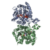


 PDBj
PDBj Assembly
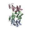
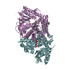


 Mass: 663.425 Da / Num. of mol.: 2 / Source method: obtained synthetically / Formula: C21H27N7O14P2 / Comment: NAD*YM
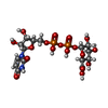
 Mass: 580.285 Da / Num. of mol.: 2 / Source method: obtained synthetically / Formula: C15H22N2O18P2
 Sample preparation
 / Beamline: X06DA / Wavelength: 1.00004 Å
 Processing