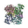
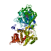
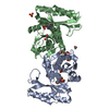
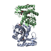
[English] 日本語
Yorodumi
- PDB-6h0n: The structure of wild-type Arabidopsis thaliana UDP-apiose/UDP-xy... -

+
Open data


ID or keywords:

Loading...

-
Basic information

Entry
Database: PDB / ID: 6h0n
TitleThe structure of wild-type Arabidopsis thaliana UDP-apiose/UDP-xylose synthase in complex with NAD+ and UDP
ComponentsUDP-D-apiose/UDP-D-xylose synthase 1
KeywordsOXIDOREDUCTASE / Enzyme catalysis / glycobiology / short-chain dehydrogenases/reductases (SDR) / aldol cleavage / substrate-assisted reaction
Function / homology
Function and homology information


nucleotide-sugar biosynthetic process / UDP-glucuronate decarboxylase activity / cell wall organization / peroxisome / NAD binding / cytoplasm / cytosol
Similarity search - Function
Bifunctional polymyxin resistance protein ArnA-like, extended (e) SDRs / NAD-dependent epimerase/dehydratase / NAD dependent epimerase/dehydratase family / NAD(P)-binding Rossmann-like Domain / NAD(P)-binding domain superfamily / Rossmann fold / 3-Layer(aba) Sandwich / Alpha Beta
Similarity search - Domain/homology
NICOTINAMIDE-ADENINE-DINUCLEOTIDE / PHOSPHATE ION / URIDINE-5'-DIPHOSPHATE / UDP-D-apiose/UDP-D-xylose synthase 1
Similarity search - Component
Biological speciesArabidopsis thaliana (thale cress)
MethodX-RAY DIFFRACTION / SYNCHROTRON / MOLECULAR REPLACEMENT / Resolution: 3.02 Å
AuthorsSavino, S. / Mattevi, A.
CitationJournal: Nat Catal / Year: 2019
Title: Deciphering the enzymatic mechanism of sugar ring contraction in UDP-apiose biosynthesis.
Authors: Savino, S. / Borg, A.J.E. / Dennig, A. / Pfeiffer, M. / de Giorgi, F. / Weber, H. / Dubey, K.D. / Rovira, C. / Mattevi, A. / Nidetzky, B.
History
DepositionJul 10, 2018Deposition site: PDBE / Processing site: PDBE
Revision 1.0Oct 23, 2019Provider: repository / Type: Initial release
Revision 1.1May 6, 2020Group: Database references / Category: citation / citation_author
Item: _citation.country / _citation.journal_abbrev ..._citation.country / _citation.journal_abbrev / _citation.journal_id_CSD / _citation.journal_id_ISSN / _citation.journal_volume / _citation.page_first / _citation.page_last / _citation.pdbx_database_id_DOI / _citation.pdbx_database_id_PubMed / _citation.title / _citation.year
Revision 1.2Jan 17, 2024Group: Data collection / Database references / Refinement description
Category: chem_comp_atom / chem_comp_bond ...chem_comp_atom / chem_comp_bond / database_2 / pdbx_initial_refinement_model / struct_ncs_dom_lim
Item: _database_2.pdbx_DOI / _database_2.pdbx_database_accession ..._database_2.pdbx_DOI / _database_2.pdbx_database_accession / _struct_ncs_dom_lim.beg_auth_comp_id / _struct_ncs_dom_lim.beg_label_asym_id / _struct_ncs_dom_lim.beg_label_comp_id / _struct_ncs_dom_lim.beg_label_seq_id / _struct_ncs_dom_lim.end_auth_comp_id / _struct_ncs_dom_lim.end_label_asym_id / _struct_ncs_dom_lim.end_label_comp_id / _struct_ncs_dom_lim.end_label_seq_id

-
Structure visualization

Structure viewerMolecule:
MolmilJmol/JSmol

Downloads & links

-
Assembly

Deposited unit
A: UDP-D-apiose/UDP-D-xylose synthase 1
B: UDP-D-apiose/UDP-D-xylose synthase 1
hetero molecules


Theoretical massNumber of molelcules
Total (without water)89,7038
Polymers87,3782
Non-polymers2,3256
Water41423
1


  • Idetical with deposited unit
  • defined by author&software
  • Evidence: gel filtration
TypeNameSymmetry operationNumber
identity operation1_555x,y,z1
Buried area8170 Å2
ΔGint-77 kcal/mol
Surface area27480 Å2
MethodPISA
Unit cell
Length a, b, c (Å)146.028, 146.028, 132.845
Angle α, β, γ (deg.)90.00, 90.00, 120.00
Int Tables number154
Space group name H-MP3221
Noncrystallographic symmetry (NCS)NCS domain:
IDEns-IDDetails
11A
21B

NCS domain segments:

Component-ID: _ / Ens-ID: 1 / Beg auth comp-ID: VAL / Beg label comp-ID: VAL / End auth comp-ID: SER / End label comp-ID: SER / Refine code: _ / Auth seq-ID: 8 - 384 / Label seq-ID: 8 - 384

Dom-IDAuth asym-IDLabel asym-ID
1AA
2BB

-
Components

#1: Protein UDP-D-apiose/UDP-D-xylose synthase 1


Mass: 43688.750 Da / Num. of mol.: 2
Source method: isolated from a genetically manipulated source
Source: (gene. exp.) Arabidopsis thaliana (thale cress) / Gene: AXS1, At2g27860, F15K20.4 / Production host: Escherichia coli BL21(DE3) (bacteria) / References: UniProt: Q9ZUY6
#2: Chemical ChemComp-NAD / NICOTINAMIDE-ADENINE-DINUCLEOTIDE


Mass: 663.425 Da / Num. of mol.: 2 / Source method: obtained synthetically / Formula: C21H27N7O14P2 / Comment: NAD*YM
#3: Chemical ChemComp-UDP / URIDINE-5'-DIPHOSPHATE


Type: RNA linking / Mass: 404.161 Da / Num. of mol.: 2 / Source method: obtained synthetically / Formula: C9H14N2O12P2 / Comment: UDP*YM
#4: Chemical ChemComp-PO4 / PHOSPHATE ION


Mass: 94.971 Da / Num. of mol.: 2 / Source method: isolated from a natural source / Formula: PO4
#5: Water ChemComp-HOH / water


Mass: 18.015 Da / Num. of mol.: 23 / Source method: isolated from a natural source / Formula: H2O

-
Experimental details

-
Experiment

ExperimentMethod: X-RAY DIFFRACTION / Number of used crystals: 1

-
Sample preparation

CrystalDensity Matthews: 4.79 Å3/Da / Density % sol: 74.3 %
Crystal growTemperature: 293 K / Method: evaporation / pH: 7.5
Details: 0.8 M sodium phosphate, 0.8 M potassium phosphate, 0.1 M sodium-HEPES

-
Data collection

DiffractionMean temperature: 100 K
Diffraction sourceSource: SYNCHROTRON / Site: SLS / Beamline: X06DA / Wavelength: 1.00368 Å
DetectorType: DECTRIS PILATUS 2M-F / Detector: PIXEL / Date: Feb 27, 2017
RadiationProtocol: SINGLE WAVELENGTH / Monochromatic (M) / Laue (L): M / Scattering type: x-ray
Radiation wavelengthWavelength: 1.00368 Å / Relative weight: 1
ReflectionResolution: 3.02→126.46 Å / Num. obs: 32264 / % possible obs: 98.9 % / Redundancy: 4.4 % / CC1/2: 0.992 / Rmerge(I) obs: 0.146 / Net I/σ(I): 9.2
Reflection shellResolution: 3.02→3.18 Å / Redundancy: 4.5 % / Rmerge(I) obs: 0.8 / Mean I/σ(I) obs: 1 / Num. unique obs: 4488 / CC1/2: 0.378 / % possible all: 95.4

-
Processing

Software
NameVersionClassification
REFMAC5.8.0158refinement
XDSdata reduction
Aimlessdata scaling
PHASERphasing
RefinementMethod to determine structure: MOLECULAR REPLACEMENT
Starting model: 3SLG
Resolution: 3.02→126.46 Å / Cor.coef. Fo:Fc: 0.944 / Cor.coef. Fo:Fc free: 0.917 / SU B: 19.584 / SU ML: 0.31 / Cross valid method: THROUGHOUT / ESU R: 0.611 / ESU R Free: 0.345 / Details: HYDROGENS HAVE BEEN ADDED IN THE RIDING POSITIONS
RfactorNum. reflection% reflectionSelection details
Rfree0.25278 1640 5.1 %RANDOM
Rwork0.20711 ---
obs0.20944 30595 98.84 %-
Solvent computationIon probe radii: 0.8 Å / Shrinkage radii: 0.8 Å / VDW probe radii: 1.2 Å
Displacement parametersBiso mean: 77.855 Å2
Baniso -1Baniso -2Baniso -3
1-1.54 Å20.77 Å20 Å2
2--1.54 Å2-0 Å2
3----5.01 Å2
Refinement stepCycle: 1 / Resolution: 3.02→126.46 Å
ProteinNucleic acidLigandSolventTotal
Num. atoms5840 0 148 23 6011
Refine LS restraints
Refine-IDTypeDev idealDev ideal targetNumber
X-RAY DIFFRACTIONr_bond_refined_d0.0130.0196124
X-RAY DIFFRACTIONr_bond_other_d0.0020.025553
X-RAY DIFFRACTIONr_angle_refined_deg1.8241.9948354
X-RAY DIFFRACTIONr_angle_other_deg1.076312878
X-RAY DIFFRACTIONr_dihedral_angle_1_deg7.3635742
X-RAY DIFFRACTIONr_dihedral_angle_2_deg39.30924.111270
X-RAY DIFFRACTIONr_dihedral_angle_3_deg17.06415983
X-RAY DIFFRACTIONr_dihedral_angle_4_deg20.3811538
X-RAY DIFFRACTIONr_chiral_restr0.0990.2941
X-RAY DIFFRACTIONr_gen_planes_refined0.0080.0216713
X-RAY DIFFRACTIONr_gen_planes_other0.0020.021227
X-RAY DIFFRACTIONr_nbd_refined
X-RAY DIFFRACTIONr_nbd_other
X-RAY DIFFRACTIONr_nbtor_refined
X-RAY DIFFRACTIONr_nbtor_other
X-RAY DIFFRACTIONr_xyhbond_nbd_refined
X-RAY DIFFRACTIONr_xyhbond_nbd_other
X-RAY DIFFRACTIONr_metal_ion_refined
X-RAY DIFFRACTIONr_metal_ion_other
X-RAY DIFFRACTIONr_symmetry_vdw_refined
X-RAY DIFFRACTIONr_symmetry_vdw_other
X-RAY DIFFRACTIONr_symmetry_hbond_refined
X-RAY DIFFRACTIONr_symmetry_hbond_other
X-RAY DIFFRACTIONr_symmetry_metal_ion_refined
X-RAY DIFFRACTIONr_symmetry_metal_ion_other
X-RAY DIFFRACTIONr_mcbond_it5.4087.7862983
X-RAY DIFFRACTIONr_mcbond_other5.4077.7852982
X-RAY DIFFRACTIONr_mcangle_it7.93511.6863720
X-RAY DIFFRACTIONr_mcangle_other7.93411.6873721
X-RAY DIFFRACTIONr_scbond_it5.838.1473141
X-RAY DIFFRACTIONr_scbond_other5.838.1483140
X-RAY DIFFRACTIONr_scangle_it
X-RAY DIFFRACTIONr_scangle_other8.8512.0544635
X-RAY DIFFRACTIONr_long_range_B_refined10.78291.6956847
X-RAY DIFFRACTIONr_long_range_B_other10.78291.6886848
X-RAY DIFFRACTIONr_rigid_bond_restr
X-RAY DIFFRACTIONr_sphericity_free
X-RAY DIFFRACTIONr_sphericity_bonded
Refine LS restraints NCS

Ens-ID: 1 / Number: 23974 / Refine-ID: X-RAY DIFFRACTION / Type: interatomic distance / Rms dev position: 0.08 Å / Weight position: 0.05

Dom-IDAuth asym-ID
1A
2B
LS refinement shellResolution: 3.018→3.096 Å / Total num. of bins used: 20
RfactorNum. reflection% reflection
Rfree0.377 133 -
Rwork0.369 2057 -
obs--91.25 %

+
About Yorodumi

-
News

-
Feb 9, 2022. New format data for meta-information of EMDB entries

New format data for meta-information of EMDB entries

  • Version 3 of the EMDB header file is now the official format.
  • The previous official version 1.9 will be removed from the archive.

Related info.:EMDB header

External links:wwPDB to switch to version 3 of the EMDB data model

-
Aug 12, 2020. Covid-19 info

Covid-19 info

URL: https://pdbjlvh1.pdbj.org/emnavi/covid19.php

New page: Covid-19 featured information page in EM Navigator.

Related info.:Covid-19 info / Mar 5, 2020. Novel coronavirus structure data

+
Mar 5, 2020. Novel coronavirus structure data

Novel coronavirus structure data

Related info.:Yorodumi Speices / Aug 12, 2020. Covid-19 info

External links:COVID-19 featured content - PDBj / Molecule of the Month (242):Coronavirus Proteases

+
Jan 31, 2019. EMDB accession codes are about to change! (news from PDBe EMDB page)

EMDB accession codes are about to change! (news from PDBe EMDB page)

  • The allocation of 4 digits for EMDB accession codes will soon come to an end. Whilst these codes will remain in use, new EMDB accession codes will include an additional digit and will expand incrementally as the available range of codes is exhausted. The current 4-digit format prefixed with “EMD-” (i.e. EMD-XXXX) will advance to a 5-digit format (i.e. EMD-XXXXX), and so on. It is currently estimated that the 4-digit codes will be depleted around Spring 2019, at which point the 5-digit format will come into force.
  • The EM Navigator/Yorodumi systems omit the EMD- prefix.

Related info.:Q: What is EMD? / ID/Accession-code notation in Yorodumi/EM Navigator

External links:EMDB Accession Codes are Changing Soon! / Contact to PDBj

+
Jul 12, 2017. Major update of PDB

Major update of PDB

  • wwPDB released updated PDB data conforming to the new PDBx/mmCIF dictionary.
  • This is a major update changing the version number from 4 to 5, and with Remediation, in which all the entries are updated.
  • In this update, many items about electron microscopy experimental information are reorganized (e.g. em_software).
  • Now, EM Navigator and Yorodumi are based on the updated data.

External links:wwPDB Remediation / Enriched Model Files Conforming to OneDep Data Standards Now Available in the PDB FTP Archive

-
Yorodumi

Thousand views of thousand structures

  • Yorodumi is a browser for structure data from EMDB, PDB, SASBDB, etc.
  • This page is also the successor to EM Navigator detail page, and also detail information page/front-end page for Omokage search.
  • The word "yorodu" (or yorozu) is an old Japanese word meaning "ten thousand". "mi" (miru) is to see.

Related info.:EMDB / PDB / SASBDB / Comparison of 3 databanks / Yorodumi Search / Aug 31, 2016. New EM Navigator & Yorodumi / Yorodumi Papers / Jmol/JSmol / Function and homology information / Changes in new EM Navigator and Yorodumi

Read more